

Imaging of liver metastases: MRI
- PMID: 17293303
- PMCID: PMC1804118
- DOI: 10.1102/1470-7330.2007.0002
Imaging of liver metastases: MRI
Abstract
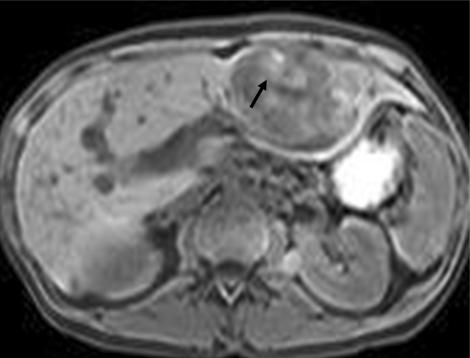
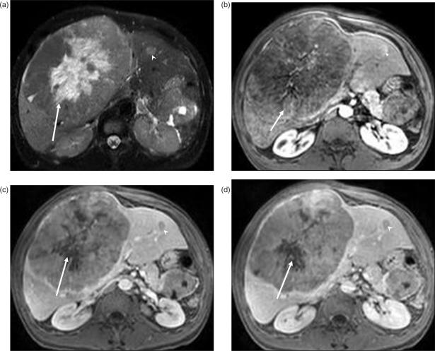
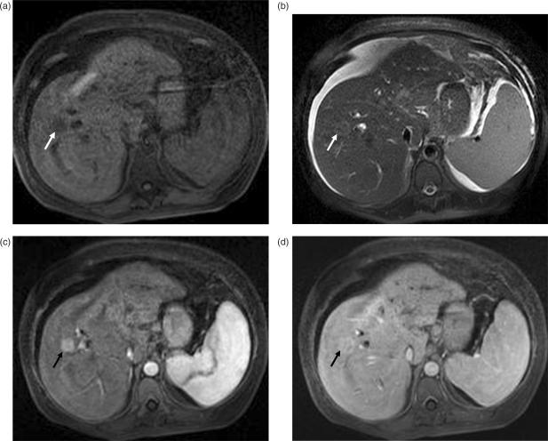
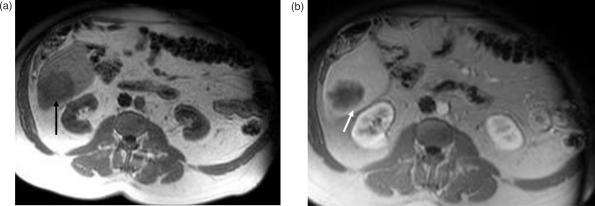
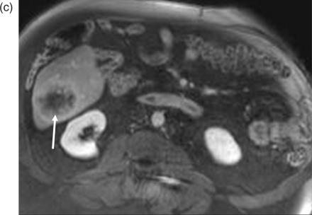
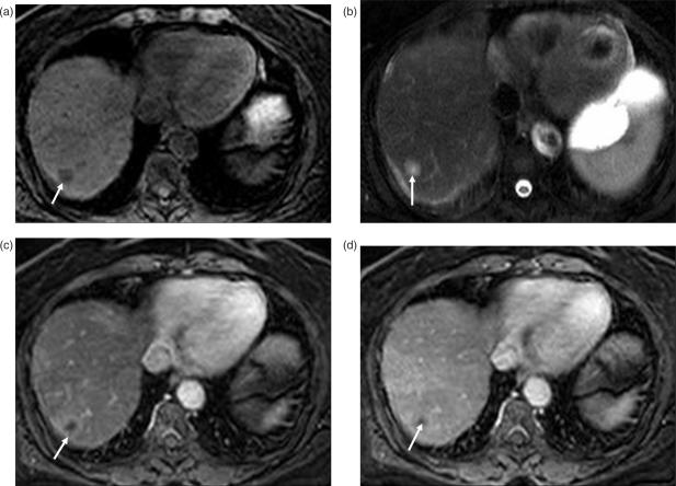
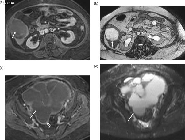
Metastases are the most common malignant liver lesions and the most common indication for hepatic imaging. Specific characterization of liver metastases in patients with primary non-hepatic tumors is crucial to avoid unnecessary diagnostic work-up for incidental benign liver lesions. Magnetic resonance (MR) is rapidly emerging as the imaging modality of choice for detection and characterization of liver lesions due to the high specificity resulting from optimal lesion-to-liver contrast and no radiation exposure. Improvements in breath-hold T1-weighted fast spoiled gradient echo and rapid T2-weighted single shot echo-train acquisition enable imaging of the liver in a single breath-hold with high spatial resolution. Most metastases are hypo- to isointense on T1 and iso- to hyperintense on T2-weighted images. MR contrast agents provide critical tumor characterization and can be safely used in patients with iodine contrast allergy and renal failure. Other agents, including newly developing gadolinium-chelates or iron oxide agents may provide additional benefits in selected applications. The degree and nature of tumor vascularity form the basis for liver lesion characterization based on enhancement properties. Liver metastases may be hypovascular or hypervascular. Colon, lung, breast and gastric carcinomas are the most common tumors causing hypovascular liver metastases, and typically show perilesional enhancement. Neuroendocrine tumors including carcinoid and islet cell tumors, renal cell carcinoma, breast, melanoma, and thyroid carcinoma are tumors most commonly causing hypervascular hepatic metastases, which may develop early enhancement with variable degrees of washout and peripheral rim enhancement.
Figures
References
-
- Imam K, Bluemke DA. MR imaging in the evaluation of hepatic metastases. Magn Reson Imaging Clin N Am. 2000;8:741–56. - PubMed
-
- Rummeny EJ, Marchal G. Liver imaging. Clinical applications and future perspectives. Acta Radiol. 1997;38:626–30. - PubMed
-
- Semelka RC, Cance WG, Marcos HB, Mauro MA. Liver metastases: comparison of current MR techniques and spiral CT during arterial portography for detection in 20 surgically staged cases. Radiology. 1999;213:86–91. - PubMed
-
- Soyer P, Levesque M, Caudron C, Elias D, Zeitoun G, Roche A. MRI of liver metastases from colorectal cancer vs. CT during arterial portography. J Comput Assist Tomogr. 1993;17:67–74. - PubMed
Publication types
MeSH terms
Substances
LinkOut - more resources
Full Text Sources
Medical