. 2007 May;189(9):3547-55.
doi: 10.1128/JB.00093-07. Epub 2007 Feb 9.

Assembly of fimbrial structures in Pseudomonas aeruginosa: functionality and specificity of chaperone-usher machineries

Affiliations

Assembly of fimbrial structures in Pseudomonas aeruginosa: functionality and specificity of chaperone-usher machineries

Ségolène Ruer et al. J Bacteriol. 2007 May.

Abstract

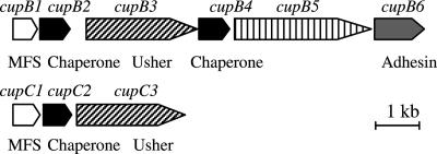
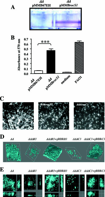
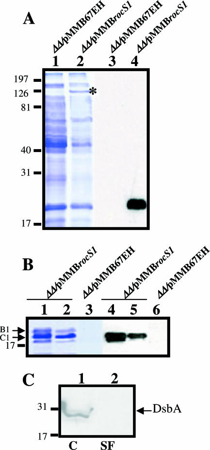
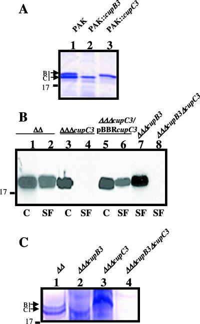
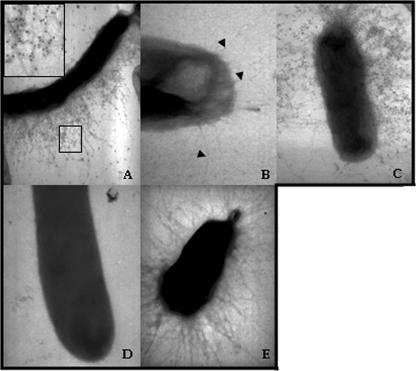
Fimbrial or nonfimbrial adhesins assembled by the bacterial chaperone-usher pathway have been demonstrated to play a key role in pathogenesis. Such an assembly mechanism has been exemplified in uropathogenic Escherichia coli strains with the Pap and the Fim systems. In Pseudomonas aeruginosa, three gene clusters (cupA, cupB, and cupC) encoding chaperone-usher pathway components have been identified in the genome sequence of the PAO1 strain. The Cup systems differ from the Pap or Fim systems, since they obviously lack numbers of genes encoding fimbrial subunits. Nevertheless, the CupA system has been demonstrated to be involved in biofilm formation on solid surfaces, whereas the role of the CupB and CupC systems in biofilm formation could not be clearly elucidated. Moreover, these gene clusters were described as poorly expressed under standard laboratory conditions. The cupB and cupC clusters are directly under the control of a two-component regulatory system designated RocA1/S1/R. In this study, we revealed that Roc1-dependent induction of the cupB and cupC genes resulted in a high level of biofilm formation, with CupB and CupC acting with synergy in clustering bacteria for microcolony formation. Very importantly, this phenotype was associated with the assembly of cell surface fimbriae visualized by electron microscopy. Finally, we observed that the CupB and CupC systems are specialized in the assembly of their own fimbrial subunits and are not exchangeable.


Figures

FIG. 1.
FIG. 1.
Genetic organization of the P. aeruginosa cupB and cupC gene clusters. The cup genes are represented with pointed ends indicating the transcriptional orientation. Genes with identical functions are represented with similarly sized and shaded symbols. MFS, major fimbrial subunit.
FIG. 2.
FIG. 2.
Biofilm formation by P. aeruginosa upon RocS1 overproduction. Strains PAO1ΔpilAΔfliC/pMMBrocS1 and PAO1ΔpilAΔfliC/pMMB67EH were inoculated into microtiter plates. (A) After ITPG induction for 48 h at 30°C, bacterial rings that formed at the air-liquid interface were observed by crystal violet staining. (B) The amount of bacteria was quantified after extraction of crystal violet and OD570 measurements. Wells inoculated with sterile medium and with the PAO1 strain were also included as controls. ***, P < 0.001. (C) Biofilm formation at the air-liquid interface of glass slides immersed in culture medium was analyzed with the PAO1ΔpilAΔfliC/pMMBrocS1 (ΔΔ), PAO1ΔpilAΔfliCΔcupB3/pMMBrocS1 (ΔΔΔcupB3), and PAO1ΔpilAΔfliCΔcupB3/pMMBrocS1 (ΔΔΔcupC3) strains using DAPI staining and epifluorescence microscopic observation at a ×63 magnification. (D) Stacked confocal scanning laser microscopy images of biofilms of the PAO1ΔpilAΔfliC/pMMBrocS1 (ΔΔ), PAO1ΔpilAΔfliCΔcupB3/pMMBrocS1 (ΔΔΔcupB3), and PAO1ΔpilAΔfliCΔcupB3/pMMBrocS1 (ΔΔΔcupC3) strains and of the trans-complemented strains with the cupB3 (ΔΔΔcupB3 + pBBRB3) and cupC3 (ΔΔΔcupC3 + pBBRC3) genes. DAPI was used for staining, and confocal-microscopic observation was done at a ×180 magnification using z slices of 300 nm. (E) Corresponding extracted z images and their respective xy and xz planes showing biofilms formed by the PAO1ΔpilAΔfliC/pMMBrocS1 (ΔΔ), PAO1ΔpilAΔfliCΔcupB3/pMMBrocS1 (ΔΔΔcupB3), and PAO1ΔpilAΔfliCΔcupB3/pMMBrocS1 (ΔΔΔcupC3) strains and in the trans-complemented strains with the cupB3 (ΔΔΔcupB3 + pBBRB3) and cupC3 (ΔΔΔcupC3 + pBBRC3) genes.
FIG. 3.
FIG. 3.
Detection of the CupC1 subunit in whole cells and in sheared appendages. (A) Whole-cell extracts from the PAO1ΔpilAΔfliC (ΔΔ) strain containing either pMMB67EH or pMMBrocS1 were collected after 72 h of growth onto M63 plates supplemented with 0.4% l-arginine and 1 mM IPTG. The proteins were separated on 12% acrylamide-SDS gels stained with Coomassie blue (lanes 1 and 2) or transferred onto nitrocellulose and revealed with CupC1 antibodies (lanes 3 and 4). The position of the RocS1 band is indicated by an asterisk. Numbers on the left are molecular mass standards (kDa). (B) Production and detection of the CupC1 protein in SFs obtained from the bacterial cell surfaces of strains PAO1ΔpilAΔfliC/pMMBrocS1 (lanes 1, 2, 4, and 5) and PAO1ΔpilAΔfliC/pMMB67EH (lanes 3 and 6). The proteins contained in the SFs were precipitated with 50% AS (lanes 1, 3, 4, and 6) and 55% AS (lanes 2 and 5). The CupB1 and the CupC1 proteins are indicated by the upper (B1) and the lower (C1) arrows. The Coomassie blue-stained gel corresponds to lanes 1 through 3, and the blot revealed with CupC1 antibodies corresponds to lanes 4 through 6. (C) The absence of intracellular proteins in the SFs was assessed by immunodetection of the DsbA periplasmic protein using appropriate antibody. C, whole-cell extracts.
FIG. 4.
FIG. 4.
Extracellular assembly of CupC1 and CupB1 proteins in a specific usher-dependent manner. (A) Protein samples obtained from SFs from the PAK, PAK::cupB3, and PAK::cupC3 strains transformed with pMMBrocS1. Proteins contained in the SFs were precipitated with 50% AS, separated on SDS gels, and stained with Coomassie blue. The CupB1 and the CupC1 proteins are indicated by the upper (B1) and the lower (C1) arrows, respectively. (B) Detection of the CupC1 protein in whole-cell extracts (C) (lanes 1, 3, and 5) and in SFs (lanes 2, 4, 6, 7, and 8) of PAO1ΔpilAΔfliC (ΔΔ), PAO1ΔpilAΔfliCΔcupC3 (ΔΔΔcupC3), PAO1ΔpilAΔfliCΔcupC3/pBBRcupC3 (ΔΔΔcupC3/pBBRcupC3), PAO1ΔpilAΔfliCΔcupB3 (ΔΔΔcupB3), and PAO1ΔpilAΔfliCΔcupB3ΔcupC3 (ΔΔΔcupB3ΔcupC3). All strains contained the pMMBrocS1 plasmid. (C) Coomassie blue staining of proteins contained in SFs from PAO1ΔpilAΔfliC (ΔΔ, lane 1), PAO1ΔpilAΔfliCΔcupB3 (ΔΔΔcupB3, lane 2), PAO1ΔpilAΔfliCΔcupC3 (ΔΔΔcupC3, lane 3), and PAO1ΔpilAΔfliCΔcupB3ΔcupC3 (ΔΔΔcupB3ΔcupC3, lane 4). All strains contained pMMBrocS1. The CupB1 and the CupC1 proteins are identified by the upper (B1) and the lower (C1) arrows, respectively.
FIG. 5.
FIG. 5.
Images of CupB and CupC appendages at the P. aeruginosa cell surface. Long fimbriae decorated with gold particles coupled to specific antibodies directed against CupC1 were peritrichously distributed at the cell surface of the PAO1ΔpilAΔfliC strain (A), in which RocS1 was overproduced (pMMBrocS1) (inset in upper left-hand corner, ×20,000 magnification of central boxed area). The CupC1-labeled fimbriae were absent in the mutant PAO1ΔpilAΔfliCΔcupC3/pMMBrocS1 (magnification, ×50,000) (B) but recovered when the cupC3 mutation was trans-complemented with the cupC3 gene (magnification, ×20,000) (C). The unlabeled CupB fimbriae were visualized (arrowheads) in the cupC3 mutant (B) and absent in the PAO1ΔpilAΔfliCΔcupB3ΔcupC3 mutant (D) (magnification, ×50,000) but recovered when the cupB3 mutation was trans-complemented with the cupB3 gene (magnification, ×20,000) (E).

Similar articles

Cited by

References

    1. Bäumler, A. J., R. M. Tsolis, F. A. Bowe, J. G. Kuster, S. Hoffmann, and F. Heffron. 1996. The pef fimbrial operon of Salmonella typhimurium mediates adhesion to murine small intestine and is necessary for fluid accumulation in the infant mouse. Infect. Immun. 64:61-68. - PMC - PubMed
    1. Choudhury, D., A. Thompson, V. Stojanoff, S. Langermann, J. Pinker, S. J. Hultgren, and S. D. Knight. 1999. X-ray structure of the FimC-FimH chaperone-adhesin complex from uropathogenic Escherichia coli. Science 285:1061-1066. - PubMed
    1. D'Argenio, D. A., M. W. Calfee, P. B. Rainey, and E. C. Pesci. 2002. Autolysis and autoaggregation in Pseudomonas aeruginosa colony morphology mutants. J. Bacteriol. 184:6481-6489. - PMC - PubMed
    1. de Bentzmann, S., M. Aurouze, G. Ball, and A. Filloux. 2006. FppA, a novel Pseudomonas aeruginosa prepilin peptidase involved in assembly of type IVb pili. J. Bacteriol. 188:4851-4860. - PMC - PubMed
    1. de Oliveira-Garcia, D., M. Dall'Agnol, M. Rosales, A. C. Azzuz, N. Alcantara, M. B. Martinez, and J. A. Giron. 2003. Fimbriae and adherence of Stenotrophomonas maltophilia to epithelial cells and to abiotic surfaces. Cell. Microbiol. 5:625-636. - PubMed

Publication types

MeSH terms