. 2007 Apr;143(4):1576-89.
doi: 10.1104/pp.106.092296. Epub 2007 Feb 9.

An early nodulin-like protein accumulates in the sieve element plasma membrane of Arabidopsis

Affiliations

An early nodulin-like protein accumulates in the sieve element plasma membrane of Arabidopsis

Junaid A Khan et al. Plant Physiol. 2007 Apr.

Abstract

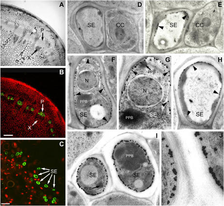
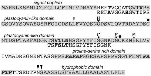
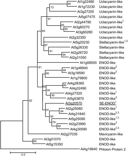
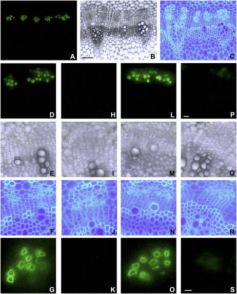
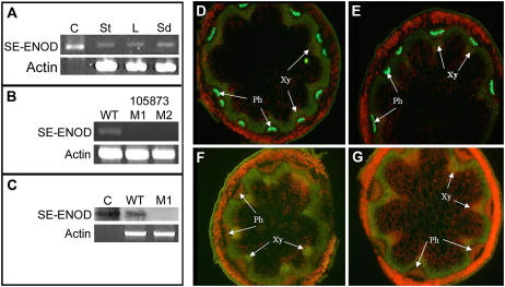
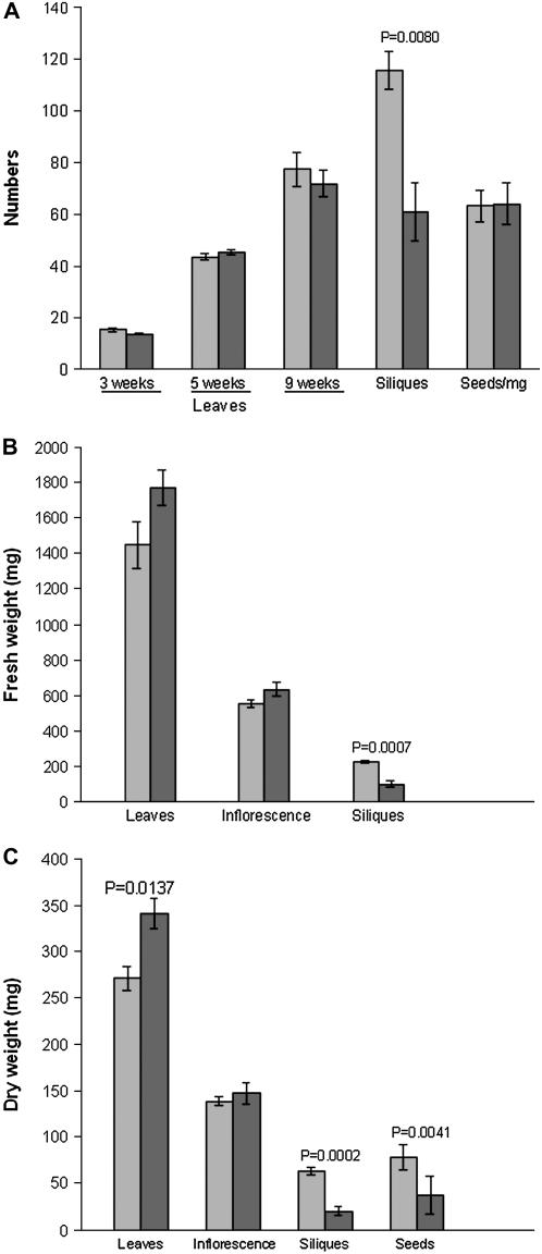
Membrane proteins within the sieve element-companion cell complex have essential roles in the physiological functioning of the phloem. The monoclonal antibody line RS6, selected from hybridomas raised against sieve elements isolated from California shield leaf (Streptanthus tortuosus; Brassicaceae) tissue cultures, recognizes an antigen in the Arabidopsis (Arabidopsis thaliana) ecotype Columbia that is associated specifically with the plasma membrane of sieve elements, but not companion cells, and accumulates at the earliest stages of sieve element differentiation. The identity of the RS6 antigen was revealed by reverse transcription-PCR of Arabidopsis leaf RNA using degenerate primers to be an early nodulin (ENOD)-like protein that is encoded by the expressed gene At3g20570. Arabidopsis ENOD-like proteins are encoded by a multigene family composed of several types of structurally related phytocyanins that have a similar overall domain structure of an amino-terminal signal peptide, plastocyanin-like copper-binding domain, proline/serine-rich domain, and carboxy-terminal hydrophobic domain. The amino- and carboxy-terminal domains of the 21.5-kD sieve element-specific ENOD are posttranslationally cleaved from the precursor protein, resulting in a mature peptide of approximately 15 kD that is attached to the sieve element plasma membrane via a carboxy-terminal glycosylphosphatidylinositol membrane anchor. Many of the Arabidopsis ENOD-like proteins accumulate in gametophytic tissues, whereas in both floral and vegetative tissues, the sieve element-specific ENOD is expressed only within the phloem. Members of the ENOD subfamily of the cupredoxin superfamily do not appear to bind copper and have unknown functions. Phenotypic analysis of homozygous T-DNA insertion mutants for the gene At3g20570 shows minimal alteration in vegetative growth but a significant reduction in the overall reproductive potential.


Figures

Figure 1.
Figure 1.
Confocal and high-resolution transmission electron microscopy immunolocalization of the SE-ENOD. A, Brightfield image showing the phloem (P) and xylem (X) in fresh tissue vibratome sections (50–100 μm) of cauliflower stem. B, The RS6 monoclonal antibody labeled only the phloem in the vascular bundles of cauliflower stem sections (bar = 200 μm). C, Immunolabeling was specific to the sieve elements (SE) of the phloem in the midrib of an Arabidopsis leaf section (bar = 10 μm). D, Transmission electron micrograph of the normal serum control treatment (no RS6 mab) shows minimal background immunogold labeling of the sieve element-companion cell (CC) complex of Arabidopsis shoot tips. E, Immunogold labeled SE-ENOD was observed only in the periphery of the sieve element and not in the companion cell. F, G, I, and J, Immunogold labeled SE-ENOD was detected in immature sieve elements, which were identified by the presence of a nucleus (N) and nondispersed P-protein bodies (PPB). H, Labeling in the mature sieve elements demonstrates that the SE-ENOD is also present in translocating sieve elements. J, High magnification of the cell wall between two immature sieve elements (see I) show the RS6 antigen localizes to the SEPM. Black arrowheads point out examples of the labeling of the plasma membrane-bound RS6 antigen with the gold particles, which can easily be observed around the entire periphery of the sieve elements.
Figure 2.
Figure 2.
The Arabidopsis At3g20570 gene encodes the 203-amino acid precursor SE-ENOD protein. The identity and arrangement of the domains (highlighted) are similar to the ENOD subclass of phyotcyanins, which is composed of an amino-terminal signal peptide, plastocyanin-like copper-binding domain, Pro-Ser-rich domain, and carboxy-terminal hydrophobic domain. The amino acid sequences below the lines show the positions of the empirically derived amino-terminal and internal California shield leaf peptide sequences. Amino acid differences between Arabidopsis and California shield leaf are indicated in bold. The gray arrowhead shows the relative position of the single intron in the codon for Leu-63 that is conserved in ENOD genes. Letters above the black inverted triangles show conserved contact residues in plastocyanins that are involved in copper binding. Black circles show the conserved Cys residues that form a disulfide linkage in phytocyanins. The two sequences (bold italics) within the Pro/Ser-rich domain are perfect (PAPAP) and imperfect (PAPTP) arabinoglycan glycosylation recognition sites. The black arrowheads indicate alternative ω attachment sites for the GPI membrane anchor.
Figure 3.
Figure 3.
GUS histochemical localization of ENOD-like gene promoter∷GUS constructs in Arabidopsis. A, Seedling of an Arabidopsis Col-0 control shows no GUS labeling in any part of the plant. B to G, Histochemical localization of GUS activity in transgenic plants that are expressing the At3g20570 promoter∷GUS construct. B, Seedling of a representative transgenic plant shows that GUS activity is limited to the vascular tissue throughout the plant. C and D, Transverse sections of a transgenic Arabidopsis leaf petiole shows that GUS activity is restricted to the phloem. E, GUS activity is limited to the vasculature of all floral parts. F, Overdevelopment of the staining reaction shows GUS activity in the vasculature of the gynoecium and the filaments of the stamens. G, Strong staining was observed at the junction of the filament and anther, but not within the anther. H and L, Histochemical localization of GUS activity in transgenic floral tissues that are expressing the related ENOD-like At4g30590 promoter∷GUS construct. H, GUS activity appeared to be observed within the gynoecium and anthers of whole flowers. GUS activity was confirmed in the developing ovules in whole tissues (I) or in sections (K) of the gynoecium as well as pollen in whole tissues (J) or in sections (L) of anthers.
Figure 4.
Figure 4.
An NJ phylogenetic tree depicting relationships among the Arabidopsis phytocyanin-like proteins. Amino acid sequences were aligned using ClustalX (Thompson et al., 1997). An unrooted NJ tree (Saitu and Nei, 1987) was constructed after corrections for a high rate of substitution using the Kimura 2-parameters distance option included in ClustalX. The branch lengths are proportional to the number of amino acid substitutions per 100 residues (bar below the tree), indicating the level of divergence among sequences, and the branch point values specify the percentage bootstrap support. The position of the SE-ENOD is highlighted. The Arabidopsis phloem lectin (At4g19840) was used as an out group in all tests. Superscript 1 indicates GPI-anchor confirmed, superscript 2 indicates expressed in pollen, and superscript 3 indicates expressed in the embryo sac.
Figure 5.
Figure 5.
SE-ENOD is GPI anchored in the SEPM. A to C, Overview (bar in B = 100 μm) of the RS6 (A) immunolocalization and structure of an unstained (B) and calcofluor white-stained (C) stem section of a cauliflower plant. D to G, Positive immunolocalization control (plus RS6 mab, D and G) and the vascular structure (E and F). H to K, Negative immunolocalization control (minus RS6 mab, H and K) and the vascular structure (I and J). L to O, Negative PIPLC treatment control (minus PIPLC, plus RS6 mab: L and O) and the vascular structure (M and N). P to S, PIPLC-treated sections (plus PIPLC, plus RS6 mab: P and S) and the vascular structure (Q and R). Bar in P = 20 μm and is representative of D to F, H to J, L to N, and P and R. Bar in S = 10 μm and is representative of G, K, and O.
Figure 6.
Figure 6.
At3g20570 transcripts and SE-ENOD protein does not accumulate in the SALK 105873 and GABI-371E08 T-DNA insertion mutants. A, RT-PCR of total RNA isolated from stem (St), leaf (L), and whole seedling (Sd) tissues from wild-type (WT) Arabidopsis Col-0 shows the amplification of a 612-bp product corresponding to the open reading frame in the cloned cDNA control (C). B, RT-PCR of total leaf RNA isolated from wild-type Arabidopsis Col-0 and two SALK 105873 homozygous mutant lines (M1 and M2) shows that the 612-bp amplicon is present in the wild type but absent in the two mutant lines. C, To increase the sensitivity of the analysis, Southern blots of the PCR products showed hybridization of the SE-ENOD cDNA probe to the cloned cDNA control and wild-type Arabidopsis Col-0, but not to the M1 mutant line. D, Immunolocalization of free-hand cross sections of fresh stem tissue from Arabidopsis Col-0 with the RS6 mab showed labeling of SE-ENOD in the phloem (Ph) but not the xylem (Xy). E, A GABI-371E08 mutant line that is heterozygous for the T-DNA insertion in the At3g20570 gene showed labeling of SE-ENOD similar to WT Col-0 plants. F and G, Neither the SALK 105873-M1 (F) nor the GABI-371E08 homozygous mutant plants showed labeling for SE-ENOD.
Figure 7.
Figure 7.
Comparative analysis of the growth and reproductive potential between wild-type Arabidopsis Col-0 and SALK 105873-M1 homozygous mutant plants. The bars in all graphs illustrate the mean ± se of the respective measurements of 16 plants in each data set. Paired Student's t test was performed for all data sets to determine the statistical significance. A, The average number of leaves at 3, 5, and 9 weeks after the appearance of the true leaves, the number of siliques, and the number of seeds per milligram of seeds. The difference in number of siliques between wild-type Col-0 and SALK 105873-M1 was significant (P = 0.0080). B, The fresh weight (milligrams) of tissues taken at 25 d after bolting showed no significant difference for the leaves and inflorescence between the wild-type Col-0 and SALK 105873-M1, whereas the fresh weight of the siliques on mutant plants was significantly (P = 0.0007) reduced. C, The dry weight (milligrams) of tissues for wild-type Col-0 and SALK 105873-M1 was determined at the end of experiment. Leaves on the mutant plants accumulated significantly (P = 0.0137) more biomass than wild-type plants, whereas no difference was observed between the dry weights of the inflorescence. The dry weights of the siliques (P = 0.0002) and seeds (P = 0.0041) showed significant differences between the two groups of plants with a reduced reproductive potential for the SALK 105873-M1 mutant.

Similar articles

Cited by

References

    1. Adman ET (1991) Copper protein structures. Adv Protein Chem 42 145–197 - PubMed
    1. Barrieu F, Chaumont F, Chrispeels MJ (1998) High expression of the tonoplast aquaporin ZmTIP1 in epidermal and conducting tissues of maize. Plant Physiol 117 1153–1163 - PMC - PubMed
    1. Bateman A, Coin L, Durbin R, Finn RD, Hollich V, Griffiths-Jones S, Khanna A, Marshall M, Moxon S, Sonnhammer EL, et al (2004) The Pfam protein families database. Nucleic Acids Res 32 D138–D141 - PMC - PubMed
    1. Becker D, Kemper E, Schell J, Masterson R (1990) New plant binary vectors with selectable markers located proximal to the left T-DNA border. Plant Mol Biol 20 1195–1197 - PubMed
    1. Bird DA, Franceschi VR, Facchini PJ (2003) A tale of three cell types: alkaloid biosynthesis is localized to sieve elements in opium poppy. Plant Cell 15 2626–2635 - PMC - PubMed

Publication types