

doi: 10.1128/AEM.02038-06.
Epub 2007 Feb 9.
Detection of low-copy-number genomic DNA sequences in individual bacterial cells by using peptide nucleic acid-assisted rolling-circle amplification and fluorescence in situ hybridization
Affiliations
- PMID: 17293504
- PMCID: PMC1855634
- DOI: 10.1128/AEM.02038-06
Item in Clipboard
Detection of low-copy-number genomic DNA sequences in individual bacterial cells by using peptide nucleic acid-assisted rolling-circle amplification and fluorescence in situ hybridization
Appl Environ Microbiol.
2007 Apr.
Abstract
An approach is proposed for in situ detection of short signature DNA sequences present in single copies per bacterial genome. The site is locally opened by peptide nucleic acids, and a circular oligonucleotide is assembled. The amplicon generated by rolling circle amplification is detected by hybridization with fluorescently labeled decorator probes.
Figures
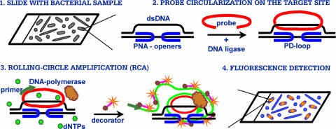
Major steps of a DNA-based assay for fluorescence in situ detection of short DNA sequences in a single bacterial cell. dNTPs, deoxynucleoside triphosphates.
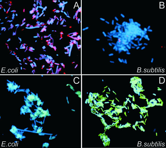
Images of bacterial cells observed by using a fluorescence microscope in experiments performed according to the scheme presented in Fig. 1. The fluorescence signals were acquired separately using three filter sets (DAPI for DNA and Cy3 or fluorescein for the labeled RCA product). Each image is a superposition of two separate images, with DAPI and Cy3 or DAPI and fluorescein signals pseudocolored in blue and red or blue and green, respectively. (A) E. coli cells to which the probes corresponding to the 21-nt target site in the E. coli cold shock protein gene (csp) region, PNA1, PNA2, ODNcspG, and decR, were applied (Table 1). Virtually all cells displayed very bright spots. No such spots were observed in numerous negative control experiments in which any of the steps of the protocol given in Fig. 1 were omitted (see the supplemental material). (B) The same procedure as that described in the legend to panel A was carried out with a combination of all probes specific to E. coli (PNA1, PNA2, PNA3, PNA4, ODNcspG, ODNrpoN, ODNrnr, and decR) (Table 1) applied to B. subtilis cells. No signal was detected. (C) A combination of probes specific to B. subtilis (PNA4, PNA5, PNA6, PNA7, ODNserA, ODNyxjA, and decG) was applied to E. coli cells. No signal was detected. (D) B. subtilis cells to which the probes corresponding to the 23-nt target site in the B. subtilis phosphoglycerate dehydrogenase gene (serA) region (PNA5, PNA6, ODNserA, and decG) were applied.
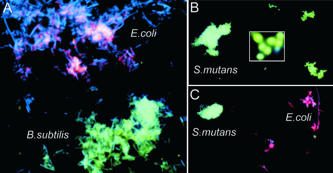
(A) Images of bacterial cells observed by using a fluorescence microscope in experiments performed with a mixture of E. coli and B. subtilis cells to which a combination of probes specific to E. coli and B. subtilis, PNA3, PNA4, PNA7, ODNrnr, ODNyxjA, decR, and decG, were applied. E. coli and B. subtilis bacteria can be distinguished from each other in the mixture of both types of cells. (B) S. mutans cells to which the probes corresponding to the 22-nt target site in the S. mutans wall-associated protein gene (wapA), PNA8, PNA9, ODNwapA, and decG, were applied. Virtually all cells are colored green. No such spots were observed in various negative control experiments in which any of the steps were omitted (data not shown). The insert shows an enlarged image of several S. mutans cells. (C) Images of bacterial cells observed by using a fluorescence microscope in experiments performed with E. coli and S. mutans cells with the combination of probes specific to E. coli (rpoN and rnr) and S. mutans (hypP): PNA3, PNA4, PNA6, ODNrpoN, ODNrnr, ODNhypP, decR, and decG. The fluorescent signals were acquired separately using three filter sets with DAPI, Cy3, and fluorescein.
Similar articles
-
Application of PNA openers for fluorescence-based detection of bacterial DNA.Methods Mol Biol. 2013;1039:223-31. doi: 10.1007/978-1-62703-535-4_18. Methods Mol Biol. 2013. PMID: 24026699
-
PNA openers and their applications for bacterial DNA diagnostics.Methods Mol Biol. 2014;1050:121-30. doi: 10.1007/978-1-62703-553-8_10. Methods Mol Biol. 2014. PMID: 24297355
-
Detection of short repeated genomic sequences on metaphase chromosomes using padlock probes and target primed rolling circle DNA synthesis.BMC Mol Biol. 2007 Nov 13;8:103. doi: 10.1186/1471-2199-8-103. BMC Mol Biol. 2007. PMID: 17997865 Free PMC article.
-
A practical approach to FRET-based PNA fluorescence in situ hybridization.Methods. 2010 Dec;52(4):343-51. doi: 10.1016/j.ymeth.2010.07.010. Epub 2010 Jul 21. Methods. 2010. PMID: 20654719 Review.
-
Lock and roll: single-molecule genotyping in situ using padlock probes and rolling-circle amplification.Histochem Cell Biol. 2006 Aug;126(2):159-64. doi: 10.1007/s00418-006-0213-2. Epub 2006 Jun 29. Histochem Cell Biol. 2006. PMID: 16807721 Review.
Cited by
-
Labeling of unique sequences in double-stranded DNA at sites of vicinal nicks generated by nicking endonucleases.Nucleic Acids Res. 2008 Apr;36(7):e40. doi: 10.1093/nar/gkn107. Epub 2008 Mar 15. Nucleic Acids Res. 2008. PMID: 18344522 Free PMC article.
-
Next-generation bis-locked nucleic acids with stacking linker and 2'-glycylamino-LNA show enhanced DNA invasion into supercoiled duplexes.Nucleic Acids Res. 2016 Mar 18;44(5):2007-19. doi: 10.1093/nar/gkw021. Epub 2016 Feb 8. Nucleic Acids Res. 2016. PMID: 26857548 Free PMC article.
-
Dual functional Phi29 DNA polymerase-triggered exponential rolling circle amplification for sequence-specific detection of target DNA embedded in long-stranded genomic DNA.Sci Rep. 2017 Jul 24;7(1):6263. doi: 10.1038/s41598-017-06594-1. Sci Rep. 2017. PMID: 28740223 Free PMC article.
-
Research Progress on Rolling Circle Amplification (RCA)-Based Biomedical Sensing.Pharmaceuticals (Basel). 2018 Apr 21;11(2):35. doi: 10.3390/ph11020035. Pharmaceuticals (Basel). 2018. PMID: 29690513 Free PMC article. Review.
-
Nanopore based sequence specific detection of duplex DNA for genomic profiling.Nano Lett. 2010 Feb 10;10(2):738-42. doi: 10.1021/nl100058y. Nano Lett. 2010. PMID: 20088590 Free PMC article.
References
-
- Abulencia, C. B., D. L. Wyborski, J. A. Garcia, M. Podar, W. Chen, S. H. Chang, H. W. Chang, D. Watson, E. L. Brodie, T. C. Hazen, and M. Keller. 2006. Environmental whole-genome amplification to access microbial populations in contaminated sediments. Appl. Environ. Microbiol. 72:3291-3301. - PMC - PubMed
-
- Amann, R., F.-O. Glockner, and A. Neef. 1997. Modern methods in subsurface microbiology: in situ identification of microorganisms with nucleic acid probes. FEMS Microbiol. Rev. 20:191-200.
-
- Bakermans, C., and E. L. Madsen. 2002. Detection in coal tar waste-contaminated groundwater of mRNA transcripts related to naphthalene dioxygenase by fluorescent in situ hybridization with tyramide signal amplification. J. Microbiol. Methods 50:75-84. - PubMed
Publication types
MeSH terms
Substances
Grants and funding
LinkOut - more resources
Full Text Sources
Other Literature Sources
