. 2007 Apr;73(8):2690-6.
doi: 10.1128/AEM.02649-06. Epub 2007 Feb 9.

Assessing adhesion forces of type I and type IV pili of Xylella fastidiosa bacteria by use of a microfluidic flow chamber

Affiliations

Assessing adhesion forces of type I and type IV pili of Xylella fastidiosa bacteria by use of a microfluidic flow chamber

Leonardo De La Fuente et al. Appl Environ Microbiol. 2007 Apr.

Abstract

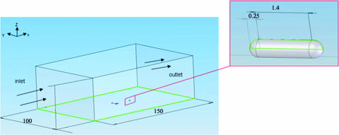
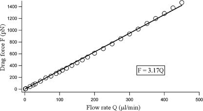
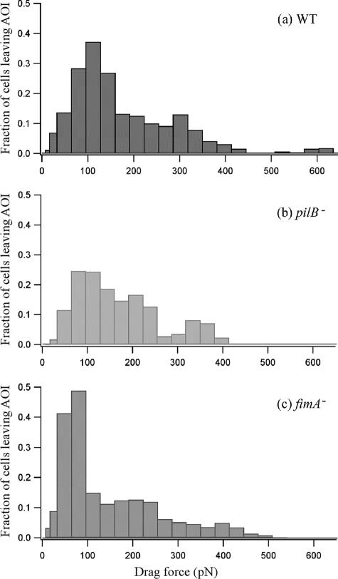
Xylella fastidiosa, a bacterium responsible for Pierce's disease in grapevines, possesses both type I and type IV pili at the same cell pole. Type IV pili facilitate twitching motility, and type I pili are involved in biofilm development. The adhesiveness of the bacteria and the roles of the two pili types in attachment to a glass substratum were evaluated using a microfluidic flow chamber in conjunction with pilus-defective mutants. The average adhesion force necessary to detach wild-type X. fastidiosa cells was 147 +/- 11 pN. Mutant cells possessing only type I pili required a force of 204 +/- 22 pN for removal, whereas cells possessing only type IV pili required 119 +/- 8 pN to dislodge these cells. The experimental results demonstrate that microfluidic flow chambers are useful and convenient tools for assessing the drag forces necessary for detaching bacterial cells and that with specific pilus mutants, the role of the pilus type can be further assessed.


Figures

FIG. 1.
FIG. 1.
Illustration of Xylella fastidiosa strains with associated pili. The WT Temecula isolate possess both the shorter type I and longer type IV pili. fimA null cells (fimA) (mutant 6E11) possess only type IV pili, and pilB null cells (pilB) possess only type I pili.
FIG. 2.
FIG. 2.
Schematic of the basic microfluidic chamber design. Microfluidic channels were 80 μm wide, 50 μm in depth, and 3.7 cm in length.
FIG. 3.
FIG. 3.
Schematic illustration of the experimental design used for numerical computation. The flow field was simulated using Fluent computation fluid dynamics software. The simulation box was 100 μm wide, 50 μm deep, and 150 μm long. The model bacterium attached to the channel surface was 0.5 μm in diameter and 1.90 μm in length.
FIG. 4.
FIG. 4.
Calibration curve generated by varying the flow rate in a test channel with total drag force exerted on the model bacterium (Fig. 3). Data points result from numerical calculations; the line represents a fit to a linear function.
FIG. 5.
FIG. 5.
Image sequences depicting detachment of Xylella fastidiosa wild-type Temecula isolate cells (WT), fimA null cells (fimA) (type IV pili only), and pilB null cells (pilB) (type I pili only) from the microfluidic chamber surface. The results for three cells are demarcated (in a rectangle, an oval, and a circle) for each of the three cell types (with one cell type shown per column) in the frames in the first row to indicate the results seen at a flow rate of 2 μl min−1 (or at a shear stress of 8 dyn/cm2). At 90 μl min−1 (or at a shear stress of 360 dyn/cm2) one each of the WT and fimA null cells (oval) detached, and all three of the mutant pilB null cells were dragged downstream. At 220 μl min−1 (or at a shear stress of 880 dyn/cm2) few WT and no fimA null cells remained attached whereas significantly more pilB null cells remained attached, albeit displaced in position downstream. Flow direction is represented from left to right.
FIG. 6.
FIG. 6.
Numbers of Xylella fastidiosa cells adherent to the microfluidic chamber surface as a function of the flow rate. Data represent the results observed for X. fastidiosa WT cells, fimA null cells (fimA), and pilB null cells (pilB).
FIG. 7.
FIG. 7.
Fraction of the cells detached from the microfluidic chamber surface as a function of the drag force. The fraction of cells is defined as follows: fraction = [(number of cells at a given flow rate − number of cells at a lower consecutive flow rate)/number of cells at the lowest flow rate in the middle two-thirds of the channel]. The plot is derived from the data presented in Fig. 6. The drag force was calculated using the relation F = 3.80Q for channel width 80 μm and F = 3.17Q for channel width 100 μm, where Q is the flow rate. fimA, fimA null cells; pilB, pilB null cells.
FIG. 8.
FIG. 8.
Adhesion force of Xylella fastidiosa WT strain cells and pilus-defective mutants (fimA, fimA null cells; pilB, pilB null cells). The different letters on the bars correspond to statistical significant differences according to pair-wise trimmed t test results (P = 0.05). Error bars, standard deviations.

References

    1. Batchelor, G. K. 1999. An introduction to fluid-dynamics, 2nd ed., p. 179-183. Cambridge University Press, Cambridge, United Kingdom.
    1. Brown, D. C., and R. S. Larson. 2001. Improvements to parallel plate flow chambers to reduce reagent and cellular requirements. BMC Immunol. 2:9. - PMC - PubMed
    1. Chaudhury, M. K., and G. M. Whitesides. 1991. Direct measurement of interfacial interactions between semispherical lenses and flat sheets of poly(dimethylsiloxane) and their chemical derivatives. Langmuir 7:1013-1025.
    1. Davis, M. J., W. J. French, and N. W. Schaad. 1981. Axenic culture of the bacteria associated with phony disease of peach and plum leaf scald. Curr. Microbiol. 6:309-314.
    1. Galvani, C. D., Yaxin Li, T. J. Burr, and H. C. Hoch. 2007. Twitching motility among pathogenic Xylella fastidiosa isolates and the influence of bovine serum albumin on twitching-dependent colony fringe morphology. FEMS Microbiol. Lett. 268:202-208. - PubMed

Publication types

LinkOut - more resources