. 2007;51(2):97-105.
doi: 10.1387/ijdb.062237ma.

Fate of cranial neural crest cells during craniofacial development in endothelin-A receptor-deficient mice

Affiliations

Fate of cranial neural crest cells during craniofacial development in endothelin-A receptor-deficient mice

Makoto Abe et al. Int J Dev Biol. 2007.

Abstract

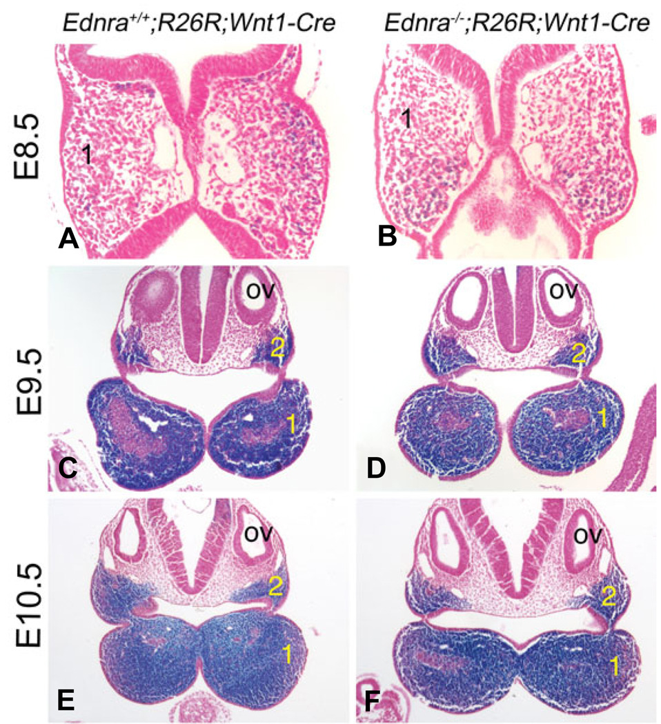
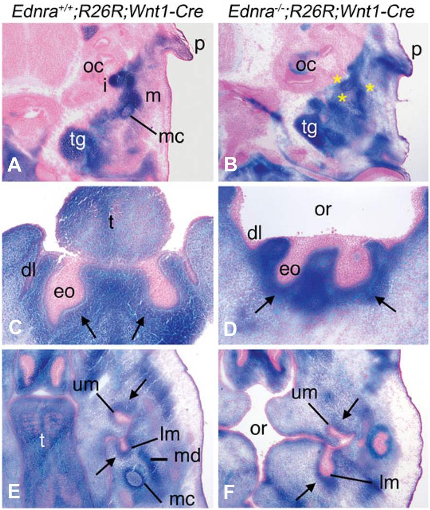
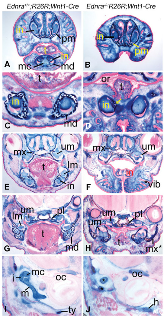
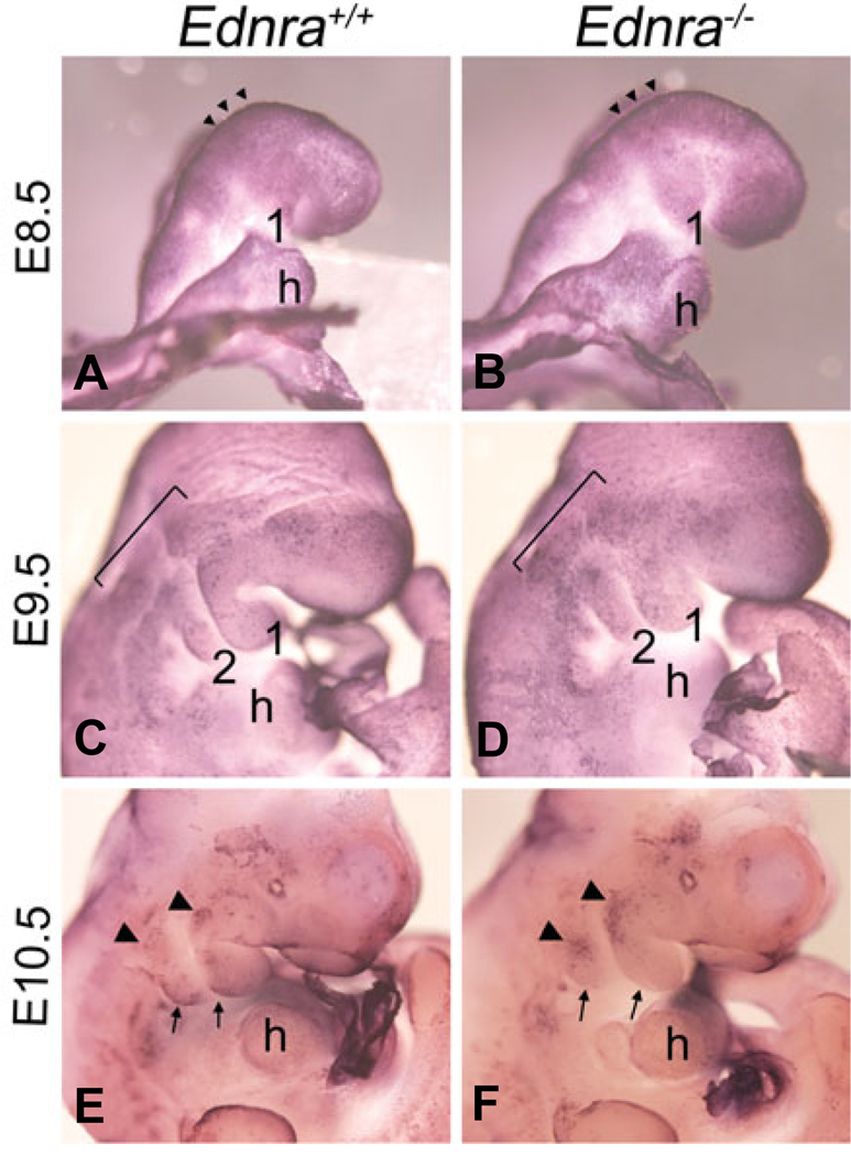
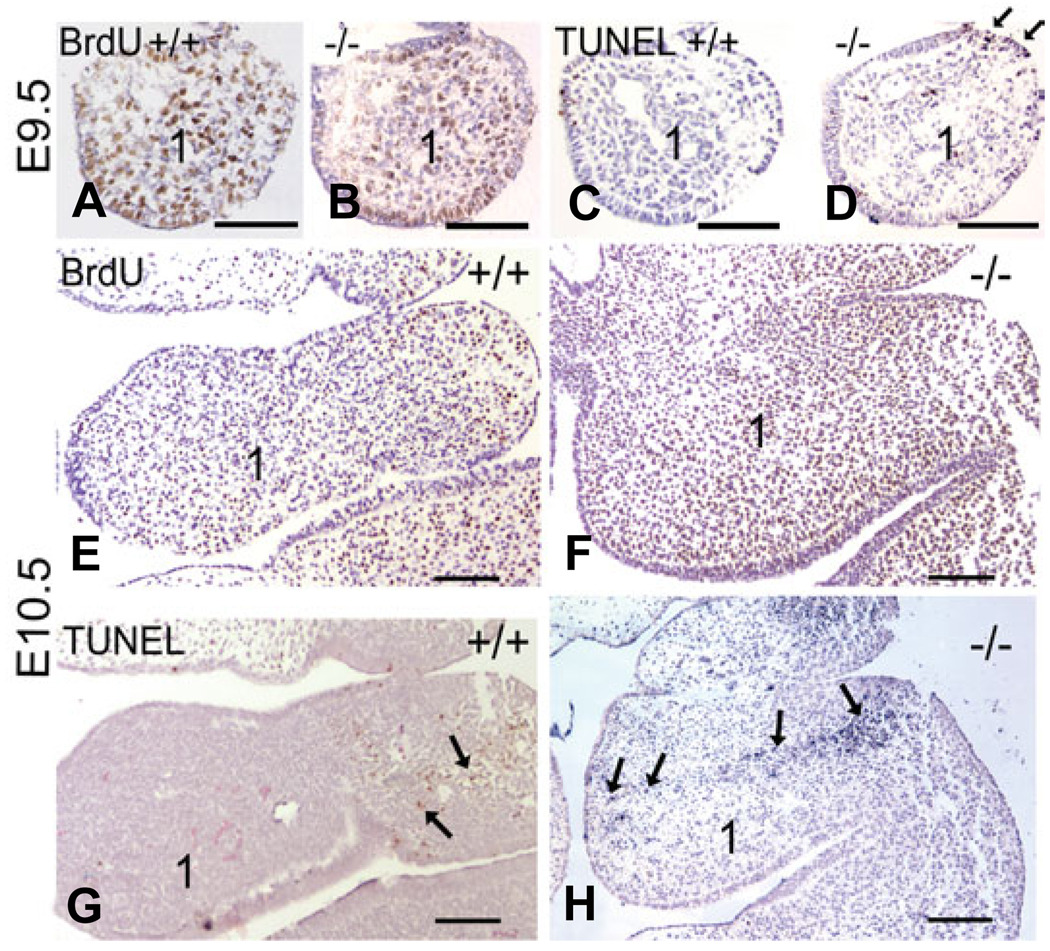
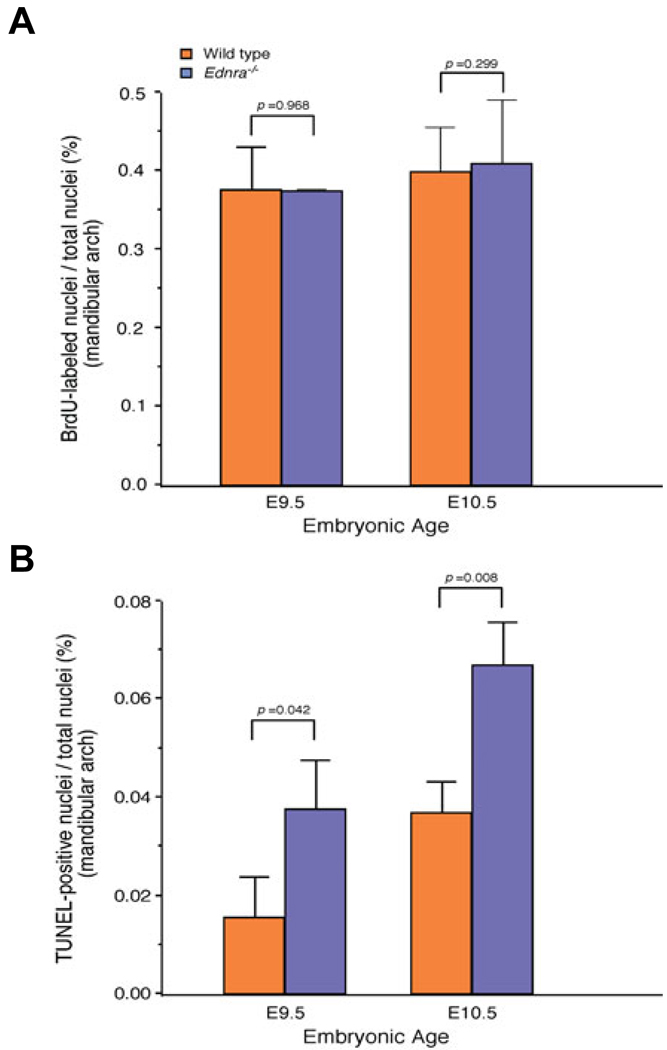
Most of the bone, cartilage and connective tissue of the lower jaw is derived from cranial neural crest cells (NCCs) arising from the posterior midbrain and hindbrain. Multiple factors direct the patterning of these NCCs, including endothelin-1-mediated endothelin A receptor (Edn1/Ednra) signaling. Loss of Ednra signaling results in multiple defects in lower jaw and neck structures, including homeotic transformation of lower jaw structures into upper jaw-like structures. However, since the Ednra gene is expressed by both migrating and post-migrating NCCs, the actual function of Ednra in cranial NCC development is not clear. Ednra signaling could be required for normal migration or guidance of NCCs to the pharyngeal arches or in subsequent events in post-migratory NCCs, including proliferation and survival. To address this question, we performed a fate analysis of cranial NCCs in Ednra-/- embryos using the R26R;Wnt1-Cre reporter system, in which Cre expression within NCCs results in permanent beta-galactosidase activity in NCCs and their derivatives. We find that loss of Ednra does not detectably alter either migration of most cranial NCCs into the mandibular first arch and second arch or their subsequent proliferation. However, mesenchymal cell apoptosis is increased two fold in both E9.5 and E10.5 Ednra-/- embryos, with apoptotic cells being present in and just proximal to the pharyngeal arches. Based on these studies, Ednra signaling appears to be required by most cranial NCCs after they reach the pharyngeal arches. However, a subset of NCCs appear to require Ednra signaling earlier, with loss of Ednra signaling likely leading to premature cessation of migration into or within the arches and subsequent cell death.


Figures

Fig. 1
Fig. 1. Localization of migratory and post-migratory NCCs in Ednra−/−;R26R;Wnt1-Cre embryos
Lateral (A–F) and ventral (E’,F’) views of Ednra+/+;R26R;Wnt1-Cre (A,C,E,E’) and Ednra−/−;R26R;Wnt1-Cre (B,D,F,F’) embryos. (A,B) At E8.5, β-gal-stained cells (blue) are observed in both Ednra+/+;R26R;Wnt1-Cre (A) and Ednra−/−;R26R;Wnt1-Cre (B) embryos extending from the neural tube toward pharyngeal arch 1 (1) and two (2), with no observed differences present between the two embryos. (C, D) At E9.5, stained cells are observed in Ednra+/+;R26R;Wnt1-Cre (C) and Ednra−/−;R26R;Wnt1-Cre (D) embryos in the frontonasal prominence, maxillary and mandibular portions of the first arch and second, third (3), fourth (4) and sixth (6) arches. No differences are observed in these regions, though stained cells in the outflow tract of wild type embryos appears to extend more distally than those in the outflow of Ednra−/−;R26R;Wnt1-Cre embryos (arrow). (E,F) The staining pattern in E10.5 embryos is similar to that observed at E9.5, with stained cells present throughout the pharyngeal arches (see also ventral views, E’ and F’). However, an apparent delay in NCC migration through the outflow tract in Ednra−/−;R26R;Wnt1-Cre embryos is still apparent (compare arrows in E and F). h, heart.
Fig. 2
Fig. 2. Histological analysis of NCCs in the pharyngeal arches of Ednra−/−;R26R;Wnt1-Cre embryos
Ednra+/+;R26R;Wnt1-Cre (A,C,E) and Ednra−/−;R26R;Wnt1-Cre (B,D,F) embryos were stained in whole mount for β-gal activity and then embedded on a transverse plane in paraffin and sectioned. Sections are counterstained with nuclear fast red. (A,B) In E8.5 embryos, labeled cells are observed in the mesenchyme of the first pharyngeal arch (1) in both Ednra+/+;R26R;Wnt1-Cre (A) and Ednra−/−;R26R;Wnt1-Cre (B) embryos. (C,D) At E9.5, most of the mesenchyme in the mandibular portion of arch one (1) and in arch two (2) in both Ednra+/+;R26R;Wnt1-Cre (C) and Ednra−/−;R26R;Wnt1-Cre (D) embryos is labeled. The unlabeled cells in the center of the arch are part of the core paraxial mesoderm. (E,F) As observed at E9.5, the arch mesenchyme of E10.5 Ednra+/+;R26R;Wnt1-Cre and Ednra−/−;R26R;Wnt1-Cre embryos is almost completely composed of labeled cells, excluding the core paraxial mesoderm. ov, otic vesicle.
Fig. 3
Fig. 3. Contribution of Ednra−/− NCCs to craniofacial structures in E13.5 Ednra−/−;R26R;Wnt1-Cre embryos
Transverse sections through the head of Ednra+/+;R26R;Wnt1-Cre (A,C,E) and Ednra−/−;R26R;Wnt1-Cre embryos (B,D,F) stained for β-gal activity. Sections were counterstained with nuclear fast red. (A,B) In sections through the lower jaw/middle ear region of Ednra+/+;R26R;Wnt1-Cre embryos, the malleus (m), incus (i) and Meckel’s cartilage (mc) contain labeled cells. Labeled cells are also observed extending toward the ear pinna (p) and in the trigeminal ganglion (tg) (A). In Ednra−/−;R26R;Wnt1-Cre embryos, labeled cells are only observed in unrecognizable cartilaginous condensations (yellow asterisks)(B). Labeled cells are also observed extending toward the pinna and in the trigeminal ganglion. (C,D) In sections through the lower incisor region, labeled cells are observed in the condensing dental mesenchyme (arrows) surrounding the enamel organ (eo) of the lower incisors and in the surrounding mesenchyme in both Ednra+/+;R26R;Wnt1-Cre (C) and Ednra−/−;R26R;Wnt1-Cre (D) embryos. (E,F) In sections through the molar region, labeled cells in Ednra+/+;R26R;Wnt1-Cre embryos compose the condensing dental mesenchyme of the developing upper and lower molars (um and lm, respectively), Meckel’s cartilage (mc) and the condensing mandible (md) (E). In Ednra−/−;R26R;Wnt1-Cre embryos, labeled cells contribute to the dental papilla of the upper and lower molars (arrow) and overall connective tissue of the head, though a definable Meckel’s cartilage and mandible are not present (F). dl, dental lamina; eo, enamel organ; oc, otic capsule; or, oral cavity; t, tongue.
Fig. 4
Fig. 4. Fate of cranial NCCs in Ednra−/−;R26R;Wnt1-Cre embryos
Frontal sections through the head of E18.5 Ednra+/+;R26R;Wnt1-Cre (A,C,E,G) and Ednra−/−;R26R;Wnt1-Cre (B,D,F,H) embryos. (A,B) Labeled cells are observed in the head mesenchyme, premaxilla (pm), mandible (md), Meckel’s cartilage (mc) and dental papilla of the upper and lower incisors (in) of Ednra+/+;R26R;Wnt1-Cre embryos (A). A similar pattern is observed in Ednra−/−;R26R;Wnt1-Cre embryos, though the recessed lower jaw is not observed (B). (C,D) In sections through the lower incisors, labeled cells are observed in the dental papilla and mandible of Ednra+/+;R26R;Wnt1-Cre embryos (C). In Ednra−/− ;R26R;Wnt1-Cre embryos (D), labeled cells are present in the dental papilla and small amount of mandible that is present (arrow). (E,F) Labeled cells in Ednra+/+;R26R;Wnt1-Cre embryos are observed in the maxilla (mx), dental papilla of upper (um) and lower (lm) molars and lower incisor (in) and surrounding mesenchyme (E). A similar pattern is observed in Ednra−/−;R26R;Wnt1-Cre embryos (F), though the lower molars are proximal to this plane of section due to the mandibular hypoplasia. (G,H) In sections proximal to those in (E,F), labeled cells in wild type embryos are observed in the mandible and palatine bones (pl), dental papilla of the upper and lower molars and surrounding mesenchyme (G). In Ednra−/−;R26R;Wnt1-Cre embryos, a similar contribution of labeled cells is observed, including the dysmorphic bone in the lower jaw that has previously been shown to have undergone homeosis to a maxilla-like structure (pseudo-maxilla, mx*). (I,J) In Ednra+/+;R26R;Wnt1-Cre embryos, the malleus (m), incus (i), Meckel’s cartilage (mc) and tympanic ring (ty) are all populated by labeled cells (I). In Ednra−/−;R26R;Wnt1-Cre embryos, aberrant cartilage (*) containing labeled cells have replaced the malleus and incus (J). Labeled cells are also present in the greater horns (h) of the hyoid, which are aberrantly present near the otic capsule (oc). or, oral cavity; t tongue; *vib, ectopic vibrissae.
Fig. 5
Fig. 5. Whole mount apoptosis analysis in Ednra−/− embryos
Lateral and ventral views of wild type (A,C,E) and Ednra−/− (B,D,F) embryos at E8.5 (A,B), E9.5 (C,D) and E10.5 (E,F) following whole mount TUNEL analysis, with NBT/BCIP as the substrate, which produces a blue deposit. (A,B) At E8.5, apoptosis is confined to the neural fold in the midbrain and hindbrain regions (arrowheads) of both wild type (A) and Ednra−/− (B) embryos. (C,D) In E9.5 wild type embryos, scattered apoptosis is observed proximal to and within pharyngeal arches one and two (C). In E9.5 Ednra−/− embryos, apoptosis is observed in a similar pattern to that of wild type embryos, though the qualitative level of apoptosis appears higher, especially in the arches (D). (E,F) In the pharyngeal arch region of E10.5 wild type embryos, apoptosis is observed in cells at the proximal aspect of arches one and two (arrowheads) and in cells along the caudal aspects of both arches (arrows) (E). In E10.5 Ednra−/− embryos, apoptosis is more prominent in the proximal aspects of the mandibular and second arches (arrowheads) but is not apparent in the caudal aspects of arches one and two (arrows) (F). h, heart.
Fig. 6
Fig. 6. Comparison of proliferation and cell death in Ednra−/− embryos
Transverse sections through the mandibular arch one and arch two of wild type (A,C,E,G) and Ednra−/− (B,D,F,H) following BrdU (A,B,E,G) or TUNEL (C,D,F,H) analysis. (A,B) BrdU labeling in the mandibular arch (1) appears similar between wild type (A) and Ednra−/− (B) embryos. (C,D) In consecutive sections to those in (A,B), few TUNEL-positive cells are observed in the mesenchyme of E9.5 wild type embryos (C), though numerous labeled nuclei (red) are observed in the mesenchyme of Ednra−/− embryos (D). There is also a local increase in TUNEL-positive cells in the ectoderm of the proximal arch (arrows in D). (E,F) At E10.5, BrdU staining appears similar between wild type (E) and Ednra−/− (F) embryos in the first arch. (G,H) In consecutive sections to those in (E,F), TUNEL-positive cells (red) in wild type embryos are confined to the proximal aspects of arches one and two (arrows in G). In Ednra−/− embryos, TUNEL-positive cells (blue) extend from this proximal region into the distal arch as denoted by arrows (H; the difference in color between labeled cells in G and H is due to different chromagens. This does not affect the outcome or sensitivity of the experiments). The magnification bar in each panel represents 100 µm.
Fig. 7
Fig. 7. Quantification of proliferation and cell death in Ednra−/− embryos
Incidence of proliferating (A) and apoptotic (B) mesenchymal cells in the mandibular arch of wild type (orange) and Ednra−/− (blue) embryos at both E9.5 and E10.5. Values represent the number of labeled nuclei as a percentage of total nuclei. p values comparing both genotypes are listed above each embryonic age.

References

    1. Bronner-Fraser M. Origins and developmental potential of the neural crest. Exp. Cell Res. 1995;218:405–417. - PubMed
    1. Chai Y, Jiang X, Ito Y, Bringas P, Han J, Rositch DH, Soriano P, Mcmahon AP, Sucov HM. Fate of the mammalian cranial neural crest during tooth and mandibular morphogenesis. Development. 2000;127:1671–1679. - PubMed
    1. Clouthier DE, Hosoda K, Richardson JA, Williams SC, Yanagisawa H, Kuwaki T, Kumada M, Hammer RE, Yanagisawa M. Cranial and cardiac neural crest defects in endothelin-A receptor-deficient mice. Development. 1998;125:813–824. - PubMed
    1. Clouthier DE, Schilling TF. Understanding endothelin-1 function during craniofacial development in the mouse and zebrafish. Birth Defects Res. (Part C) 2004;72:190–199. - PubMed
    1. Clouthier DE, Williams SC, Hammer RE, Richardson JA, Yanagisawa H. Cell-autonomous and non-autonomous actions of endothelin-A receptor signaling in craniofacial and cardiovascular development. Dev. Biol. 2003;261:506–519. - PubMed

Publication types

Substances