. 2007 May 15;404(1):31-43.
doi: 10.1042/BJ20061483.

Neurogranin binds to phosphatidic acid and associates to cellular membranes

Affiliations

Neurogranin binds to phosphatidic acid and associates to cellular membranes

Irene Domínguez-González et al. Biochem J. .

Abstract

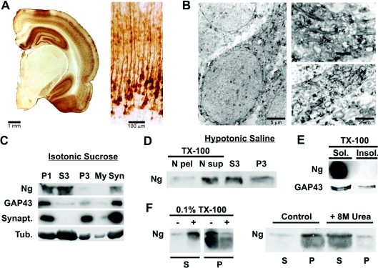
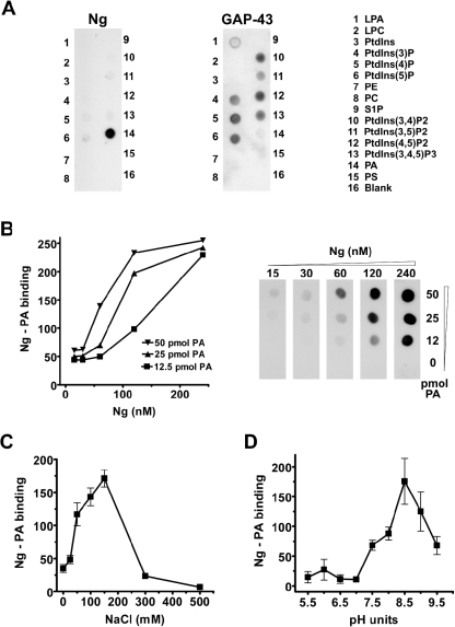
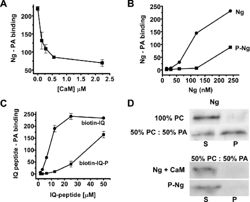
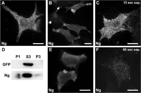
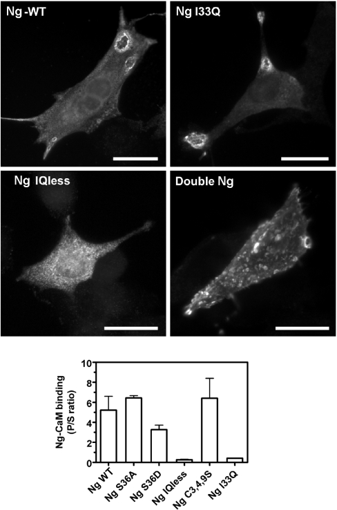
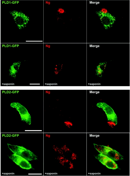
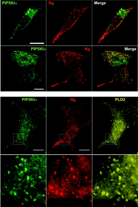
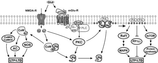
Neurogranin (Ng) is a 78-amino-acid-long protein concentrated at dendritic spines of forebrain neurons that is involved in synaptic plasticity through the regulation of CaM (calmodulin)-mediated signalling. Ng features a central IQ motif that mediates binding to CaM and is phosphorylated by PKC (protein kinase C). We have analysed the subcellular distribution of Ng and found that it associates to cellular membranes in rat brain. In vitro binding assays revealed that Ng selectively binds to PA (phosphatidic acid) and that this interaction is prevented by CaM and PKC phosphorylation. Using the peptide Ng-(29-47) and a mutant with an internal deletion (Ng-IQless), we have shown that Ng binding to PA and to cellular membranes is mediated by its IQ motif. Ng expressed in NIH-3T3 cells accumulates at peripheral regions of the plasma membrane and localizes at intracellular vesicles that can be clearly visualized following saponin permeabilization. This distribution was affected by PLD (phospholipase D) and PIP5K (phosphatidylinositol 4-phosphate 5-kinase) overexpression. Based on these results, we propose that Ng binding to PA may be involved in Ng accumulation at dendritic spines and that Ng could modulate PA signalling in the postsynaptic environment.


Figures

Figure 1
Figure 1. Ng is associated to neuronal membranes in rat brain
Brains from young adult Wistar rats were processed for immunocytochemistry as described in the Experimental section. (A) Left: stereomicroscope print showing half a coronal section displaying abundant Ng staining at the hippocampus, cerebral cortex and amygdala and none in the thalamus. Right: bright-field print showing Ng immunostaining distribution across somatosensory cortical layers. (B) Electron microscopy prints showing typical Ng immunostaining of cortical pyramidal neurons. Ng is distributed as small aggregates along an apical dendrite and frequently labels the cytoplasmic face of intracellular membranes. (C) Brains were density-fractionated in isotonic sucrose and processed by Western blotting using specific antibodies. Synapt., synaptophysin; Tub., tubulin. P1, S3, P3, My and Syn are described in the Experimental section. (D) Brains were density-fractionated in hypotonic saline and processed by Western blotting. Most of the Ng present at the P1 fraction was recovered in a soluble form after Triton X-100 (TX-100) extraction. N pel, N sup, S3 and P3 are described in the Experimental section. (E) Triton X-100 (TX-100)-insoluble (Insol.) (lipid rafts) and -soluble (Sol.) fractions were analysed by Western blotting. No Ng could be detected in the lipid raft fraction, whereas GAP-43 was clearly partitioned between Triton X-100-soluble and -insoluble fractions. (F) Synaptosomes from sucrose density fractionation were washed with isotonic sucrose and homogenized in 20 ml of a solution containing 1 mM EDTA and 10 mM Mops, pH 7.4. After stirring for 10 min at 4 °C, the mixture was centrifuged at 15000 g for 10 min at 4 °C and the pellet was resuspended and frozen in the same buffer at 1.5 mg/ml total protein. For assays, aliquots of synaptosomal membranes were thawed, centrifuged, resuspended in the same buffer with different additions, incubated for 30 min at 4 °C with gentle stirring and centrifuged again. Aliquots of supernatant (S) and pellet (P) were analysed by Western blotting. TX-100, Triton X-100.
Figure 2
Figure 2. Ng binds to PA in vitro
(A) PIP strips were blocked with 3% fatty acid-free BSA and incubated overnight with recombinant Ng or GAP-43 at 0.5 μg/ml. After extensive washing with TBST, anti-GAP-43 (797) or anti-Ng (756) affinity-purified antibodies were used to analyse protein content. The experiments were performed independently at least three times with similar results. A representative blot is shown. LPA, lysophosphatidic acid; LPC, lysophosphatidylcholine; PE, phosphatidylethanolamine; S1P, sphingosine 1-phosphate. (B) Hybond-C nitrocellulose strips were spotted with different amounts of egg-yolk PA and incubated with different concentrations of Ng. Ng–PA binding was quantified by densitometry of films exposed for different lengths of time and is expressed in arbitrary units. (C) The effect of ionic strength on Ng binding to PA was analysed by changing the NaCl concentration during overnight incubations with Ng as indicated. (D) The effect of pH on Ng binding to PA was analysed using a Tris/glycine buffer during overnight incubations with Ng. Results are means±S.E.M. for at least three independent experiments.
Figure 3
Figure 3. Regulation of Ng binding to PA
(A) Ng binding to PA was analysed in protein–lipid overlays as described in the text, except that Ng was incubated previously with different amounts of CaM for 30 min at 25 °C before the mixture was added to egg-yolk PA-spotted strips. (B) Recombinant Ng was phosphorylated by PKC and separated from non-phosphorylated Ng by affinity chromatography in CaM–agarose columns. Binding of Ng (●) and phospho-Ng (■) was assayed as described in the text. (C) Binding of biotin–IQ peptide (●), corresponding to Ng-(29–47), and its phosphorylated form (biotin–IQ-P) (■) to egg-yolk PA-spotted strips was analysed using the method described for protein–lipid overlay assays, except that the primary and secondary antibodies were replaced by an incubation with HRP-labelled streptavidin (1:2000 dilution) for 1 h. Results in (A), (B) and (C) are means±S.E.M. for at least three independent experiments. (D) To analyse CaM competition, 5 μg of CaM was added to liposomes of the indicated composition along with Ng to the binding mixture. Typical Western blots representative of three independent experiments are shown. S, supernatant; P, pellet.
Figure 4
Figure 4. Ng subcellular distribution in NIH-3T3 cells
NIH-3T3 cells were transfected with Ng alone or in conjunction with GFP. (A) Typical distribution of Ng in cells processed using the conventional IF method, showing a characteristic punctate intracellular labelling. (B, E) Ng distribution in cells processed using the cool IF method. Ng accumulates at ring-shaped structures that appear to form at the cell periphery. (C, F) Ng-transfected cells were permeabilized in situ with 0.2% saponin (sap.) for 15 s (C) or 60 s (F) and then processed using the cool IF method. Note the clustered distribution of Ng-labelled granules that spread out from the cell after 15 s of permeabilization and the spotty vesicular distribution observed after 60 s. Scale bar, 25 μm. (D) Cellular extracts from Ng- and GFP-transfected cells were density-fractionated and analysed by Western blotting. Note that, while GFP was completely recovered in the S3 fraction, visible amounts of Ng were present in the P1 and P3 fractions. P1, S3 and P3 are described in the Experimental section.
Figure 5
Figure 5. Ng localization at the plasma membrane is mediated by the IQ motif
Upper panels: NIH-3T3 cells were transfected with several wild-type Ng (Ng-WT) and Ng mutants as indicated. Cells transfected with Ng and Ng-I33Q displayed a typical distribution with strongly labelled craters, whereas Ng-IQless labelling was limited to intracellular puncta. Double-Ng distribution was characterized by its abundant presence at the plasma membrane. Scale bars, 25 μm. Lower panel: binding between CaM–agarose and several Ng mutants was assayed as described in the text. The results are expressed as the ratio between bound (P) and unbound (S) Ng in each assay. Note that Ng, Ng-S36A and Ng-C3,4,9S showed a similar binding to CaM, and Ng-IQless and Ng-I33Q did not bind CaM. Ng-S36D showed an intermediate level of CaM binding. Results are means±S.E.M. for four independent experiments.
Figure 6
Figure 6. Effects of PLD overexpresion on Ng subcellular distribution
NIH-3T3 cells were transfected with Ng and either PLD1–GFP (upper panels) or PLD2–GFP (lower panels) and analysed using the cool IF method before (upper rows) or after (lower rows) 60 s of in situ permeabilization with saponin. Note that Ng labelling at the plasma membrane is restricted to a peripheral patch that showed strong fluorescence. After saponin extraction, Ng is observed in perinuclear (PLD1) or dispersed cytoplasmic (PLD2) vesicles. These vesicles were bigger than those observed without PLD overexpression. No co-localization between Ng and PLDs was observed. Scale bar, 25 μm.
Figure 7
Figure 7. Effect of PtdIns(4,5)P2 availability on Ng subcellular distribution
Upper panels: NIH-3T3 cells were transfected with Ng and PIP5KIα and their distribution was analysed using the cool IF method. In both motile (upper row) and quiescent (lower row) cells, Ng showed a predominant distribution at the plasma membrane, characterized by its presence in multiple small protrusions or blebs that displayed some co-localization with PIP5KIα. Lower panels: NIH-3T3 cells were transfected with Ng, PIP5KIα and PLD2–HA (haemagglutinin) and their distribution was analysed as described above. The expression of PIP5KIα also alters PLD2–HA localization at the plasma membrane. PIP5KIα, PLD2–HA and Ng show a typical spotty distribution, characterized by their presence in multiple small protrusions or blebs at the cell periphery. Outlines in the upper row are shown magnified in the lower row. Co-localization of P5KIα, PLD2–HA and Ng can be observed at the protrusions.
Figure 8
Figure 8. Model depicting Ng participation in postsynaptic signal transduction
Under resting conditions (low Ca2+, low PKC activity), Ng locally traps free CaM and PA. Following glutamate receptor activation [NMDA-R (N-methyl-D-aspartate receptor) and mGlu-R (metabotropic glutamate receptor)], both CaM and PA are released and participate in signal transduction pathways that favour LTP over LTD. AC, adenylylate cyclase; DAG, diacylglycerol; mTOR: mammalian target of rapamycin; NOS, nitric oxide synthase; PP1c, PP1 catalytic subunit.

Similar articles

Cited by

References

    1. Baudier J., Deloulme J. C., Van Dorsselaer A., Black D., Matthes H. W. Purification and characterization of a brain-specific protein kinase C substrate, neurogranin (p17): identification of a consensus amino acid sequence between neurogranin and neuromodulin (GAP43) that corresponds to the protein kinase C phosphorylation site and the calmodulin-binding domain. J. Biol. Chem. 1991;266:229–237. - PubMed
    1. Watson J. B., Battenberg E. F., Wong K. K., Bloom F. E., Sutcliffe J. G. Subtractive cDNA cloning of RC3, a rodent cortex-enriched mRNA encoding a novel 78 residue protein. J. Neurosci. Res. 1990;26:397–408. - PubMed
    1. Baudier J., Bronner C., Kligman D., Cole R. D. Protein kinase C substrates from bovine brain: purification and characterization of neuromodulin, a neuron-specific calmodulin-binding protein. J. Biol. Chem. 1989;264:1824–1828. - PubMed
    1. Alvarez-Bolado G., Rodriguez-Sanchez P., Tejero-Diez P., Fairen A., Diez-Guerra F. J. Neurogranin in the development of the rat telencephalon. Neuroscience. 1996;73:565–580. - PubMed
    1. Yasuda H., Barth A. L., Stellwagen D., Malenka R. C. A developmental switch in the signaling cascades for LTP induction. Nat Neurosci. 2003;6:15–16. - PubMed

Publication types