. 2007 Feb 12:7:6.
doi: 10.1186/1472-6807-7-6.

A silent H-bond can be mutationally activated for high-affinity interaction of BMP-2 and activin type IIB receptor

Affiliations

A silent H-bond can be mutationally activated for high-affinity interaction of BMP-2 and activin type IIB receptor

Dionys Weber et al. BMC Struct Biol. .

Abstract

Background: Bone morphogenetic proteins (BMPs) are key regulators in the embryonic development and postnatal tissue homeostasis in all animals. Loss of function or dysregulation of BMPs results in severe diseases or even lethality. Like transforming growth factors beta (TGF-betas), activins, growth and differentiation factors (GDFs) and other members of the TGF-beta superfamily, BMPs signal by assembling two types of serine/threonine-kinase receptor chains to form a hetero-oligomeric ligand-receptor complex. BMP ligand receptor interaction is highly promiscuous, i.e. BMPs bind more than one receptor of each subtype, and a receptor bind various ligands. The activin type II receptors are of particular interest, since they bind a large number of diverse ligands. In addition they act as high-affinity receptors for activins but are also low-affinity receptors for BMPs. ActR-II and ActR-IIB therefore represent an interesting example how affinity and specificity might be generated in a promiscuous background.

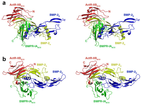
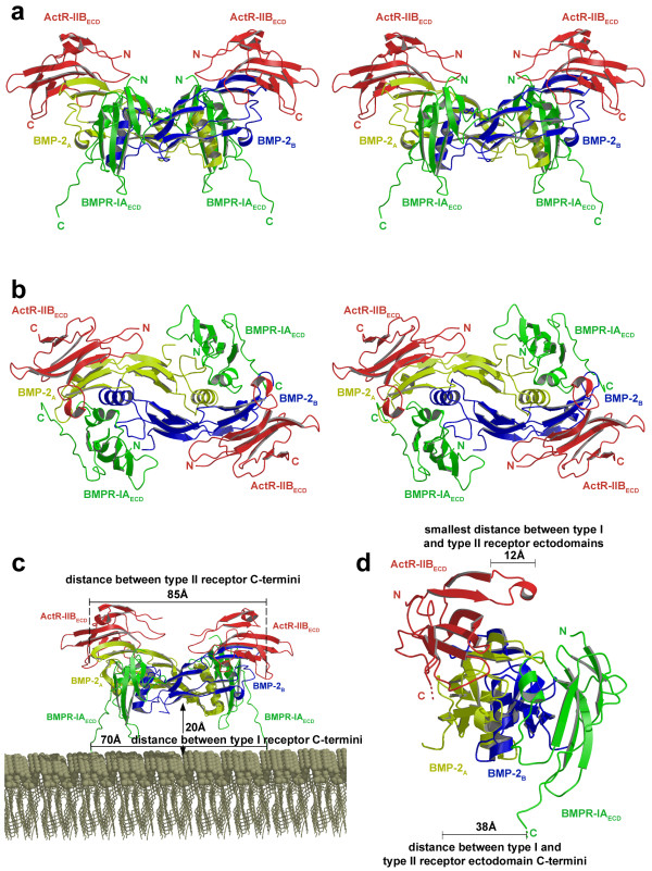
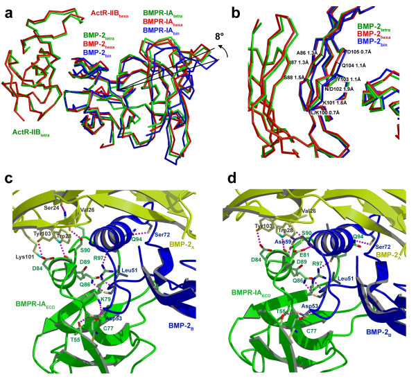
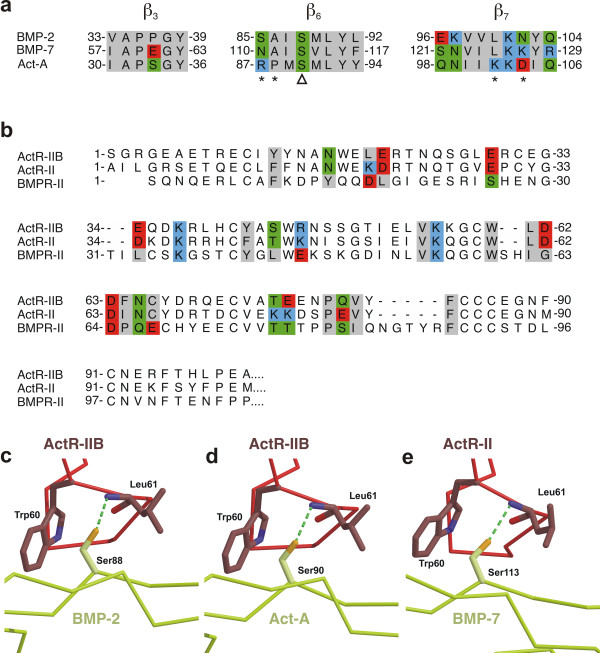
Results: Here we present the high-resolution structures of the ternary complexes of wildtype and a variant BMP-2 bound to its high-affinity type I receptor BMPR-IA and its low-affinity type II receptor ActR-IIB and compare them with the known structures of binary and ternary ligand-receptor complexes of BMP-2. In contrast to activin or TGF-beta3 no changes in the dimer architecture of the BMP-2 ligand occur upon complex formation. Functional analysis of the ActR-IIB binding epitope shows that hydrophobic interactions dominate in low-affinity binding of BMPs; polar interactions contribute only little to binding affinity. However, a conserved H-bond in the center of the type II ligand-receptor interface, which does not contribute to binding in the BMP-2 - ActR-IIB interaction can be mutationally activated resulting in a BMP-2 variant with high-affinity for ActR-IIB. Further mutagenesis studies were performed to elucidate the binding mechanism allowing us to construct BMP-2 variants with defined type II receptor binding properties.

Conclusion: Binding specificity of BMP-2 for its three type II receptors BMPR-II, Act-RII and ActR-IIB is encoded on single amino acid level. Exchange of only one or two residues results in BMP-2 variants with a dramatically altered type II receptor specificity profile, possibly allowing construction of BMP-2 variants that address a single type II receptor. The structure-/function studies presented here revealed a new mechanism, in which the energy contribution of a conserved H-bond is modulated by surrounding intramolecular interactions to achieve a switch between low- and high-affinity binding.


Figures

Figure 1
Figure 1
Ternary ligand-receptor complex of wildtype BMP-2. Ribbon representation (stereo figure) of the crystal structure of wildtype BMP-2 (monomers in yellow and blue) bound to one receptor ectodomain of BMPR-IAECD (green) and ActR-IIBECD (red), (a) viewed from the side, (b) or from above. The unexpected stoichiometry 1:1:1 is due to crystal packing forces resulting in the loss of one BMPR-IAECD and one ActR-IIBECD molecule in the ternary complex.
Figure 2
Figure 2
Ternary ligand-receptor complex of BMP-2 variant L100K/N102D. Ribbon representation (stereoview) of the ternary complex of the BMP-2 double variant L100K/N102D (in yellow and blue) bound to BMPR-IAECD (green) and ActR-IIBECD (red), viewed from the side (a) or from above (b). (c) Distances between the C-termini of the receptor ectodomains of each subtype are indicated. (d) The shortest distance between BMPR-IAECD and ActR-IIBECD occurs between the two receptor ectodomains located on the same half of the BMP-2 dimer across the β-sheet of BMP-2 and measures ~12 Å. No direct receptor-receptor contacts between the ectodomains of either subtype as proposed for the TGF-β:TGF-β receptor interaction [44, 47, 61] can be observed.
Figure 3
Figure 3
BMP-2 type I receptor interface. (a) The tilt angle of BMPR-IA bound to BMP-2 changes upon binding of the type II receptor ActR-IIB. A superposition of the structures of BMP-2:(BMPR-IAECD)2 (blue, PDB entry 1REW), the ternary complex (1:1:1) of wildtype BMP-2:BMPR-IAECD:ActR-IIBECD (green) and the ternary complex (1:2:2) of BMP-2L100K/N102D:(BMPR-IAECD)2:(ActR-IIBECD)2 (red) is shown. The comparison of both assemblies reveals that the rearrangement is not due to the mutations introduced in BMP-2L100K/N102D. (b) A change in the backbone conformation of residues 86 to 88, and 100 to 105 located in the finger 2 of BMP-2 is the possible cause for the tilt angle change. The type I ligand-receptor interfaces of the ternary (1:1:1) (c) and (1:2:2) (d) BMP-2/receptor complexes are structurally almost identical differing only in very few H-bonds that are located at the solvent accessible surface.
Figure 4
Figure 4
BMP-2 type II receptor interface. (a) Location of the type II ligand/receptor binding epitopes on wildtype BMP-2 (left) and ActR-IIBECD(right). For designation of β-strands and finger-like structures see [62], the contact residues are marked in grey. (b) Surface representation of the type II ligand/receptor binding epitopes in the ''open book'' view. The surface of BMP-2 (left) is color coded by amino acid properties as follows: hydrophobic amino acids (A, F, G, I, L, M, P, V, W, Y) are shown in white/grey, polar residues in bright/dark green (H, N, Q, S, T), acidic residues in orange/red (D, E) and positively charged amino acids (K, R) in cyan/blue. Darker colors mark the contact interface. The surface of ActR-IIBECD (right) is color coded identically. (c) Contact scheme of the wildtype BMP-2:ActR-IIBECD interaction. Intermolecular van der Waals contacts (cutoff 4.5 Å) are marked by lines, H-bonds are shown in red. Contacts involving hydrophobic residues of BMP-2 are shown in the left panel, interactions involving polar residues of BMP-2 are on the right. The surface area (Å2) buried upon complex formation is indicated by small numbers.
Figure 5
Figure 5
Binding epitopes of BMPs and activin for interaction with activin receptors are very similar. (a) Structure based sequence alignment for the regions of BMP-2, BMP-7 and Act-A building the knuckle epitope. The putative contact residues based on the BMP-2:ActR-IIB interaction are color coded according to Fig. 4b. Asterisks mark the amino acid positions chosen for „domain swapping" between BMP-2 and Act-A, the conserved Ser is indicated by a triangle. (b) Sequence alignment of the extracellular domain of ActR-IIB, ActR-II and BMPR-II, the residues contributing to the binding epitope (based on the BMP-2:ActR-IIB interface of this study) are color coded on amino acid properties as in Fig. 4b. (c-e) Comparison of the structural environment around the central H-bond in the complexes of (c) wildtype BMP-2:BMPR-IA:ActR-IIB, (d) Act-A:ActR-IIB (PDB entry 1S4Y, [42]) and (e) BMP-7:ActR-II (PDB entry 1LX5, [40]).
Figure 6
Figure 6
Mechanism for affinity switch in BMP-2 L100K/N102D. H-bond network around the conserved serine residue in Act-A (a), the BMP-2 variant L100K/N102D with increased ActR-IIB affinity (b) and wildtype BMP-2 (c). The conserved central H-bond between Ser88 Oγ (Ser90 in Act-A) and Leu61 amide of ActR-IIB is shown as green thick stippled line. The intramolecular H-bond network comprising Lys100, Asp102, Ser88 (Lys102, Asp104 and Ser90 in Act-A) and a nearby structurally conserved water molecule is indicated by stippled lines in magenta. The putative intermolecular H-bond between Lys102 of Act-A and Cys59 backbone carbonyl of ActR-IIB in the structure of the complex Act-A:ActR-IIBECD (PDB entry 1S4Y, [42]) is indicated by a thin line (a), as this H-bond is only present on one half of the dimeric complex and its geometrical parameters are close to exclusion cutoff criteria. A comparison of the position of the structurally conserved water molecule in the three structures shows that in wildtype BMP-2 (c) this solvent molecule is located directly above the central H-bond indicating direct accessibility of the H-bond by solvent. (d, e) Surface representation of ActR-IIB color coded by the contribution (ΔΔG in kJ mol-1) of each residue side chain to the binding free energy for (d) wildtype BMP-2 and (e) Act-A as measured by alanine scanning mutational analysis (see Table 1). For residue L61 the exchange to proline was used to point out the influence of the central conserved H-bond. The ΔΔG values are given in kJ mol-1. Dark red color marks hot spots of binding with an energy contribution of more than 15 kJ mol-1. Residues in yellow contribute only little; energy contribution of residues marked in blue is considered insignificant.
Figure 7
Figure 7
Biological activities of specificity-altered BMP-2 variants. (a) The BMP variants L100K/N102D (red) and S85R/A86P (green) exhibit a two- to three-fold lower EC50 value for ALP induction as wildtype BMP-2. The increased biological activity correlates with the increased affinity for ActR-IIB/ActR-II (L100K/N102D) or BMPR-II (S85R/A86P). (b) The single variant BMP-2 L100K, which has a six-fold higher affinity for ActR-IIB, shows no increased biological activity in C2C12 cells.

Similar articles

Cited by

References

    1. Hogan BL. Bone morphogenetic proteins in development. Curr Opin Genet Dev. 1996;6:432–438. doi: 10.1016/S0959-437X(96)80064-5. - DOI - PubMed
    1. Massague J. TGF-ß signal transduction. Annu Rev Biochem. 1998;67:753–791. doi: 10.1146/annurev.biochem.67.1.753. - DOI - PubMed
    1. Reddi AH. Role of morphogenetic proteins in skeletal tissue engineering and regeneration. Nat Biotechnol. 1998;16:247–252. doi: 10.1038/nbt0398-247. - DOI - PubMed
    1. Reddi AH, (ed) Bone morphogenetic proteins. Cytokine Growth Factor Rev. 2005;16:249–376. doi: 10.1016/j.cytogfr.2005.04.003. - DOI - PubMed
    1. Savage-Dunn C. Targets of TGF ß-related signaling in Caenorhabditis elegans. Cytokine Growth Factor Rev. 2001;12:305–312. doi: 10.1016/S1359-6101(01)00015-6. - DOI - PubMed

Publication types