Review
. 2007 Feb;131(2):595-607.
doi: 10.1378/chest.06.2287.

Central sleep apnea: Pathophysiology and treatment

Affiliations
Review

Central sleep apnea: Pathophysiology and treatment

Danny J Eckert et al. Chest. 2007 Feb.

Abstract

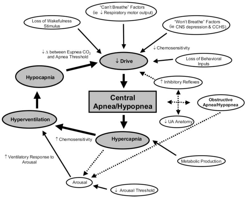
Central sleep apnea (CSA) is characterized by a lack of drive to breathe during sleep, resulting in repetitive periods of insufficient ventilation and compromised gas exchange. These nighttime breathing disturbances can lead to important comorbidity and increased risk of adverse cardiovascular outcomes. There are several manifestations of CSA, including high altitude-induced periodic breathing, idiopathic CSA, narcotic-induced central apnea, obesity hypoventilation syndrome, and Cheyne-Stokes breathing. While unstable ventilatory control during sleep is the hallmark of CSA, the pathophysiology and the prevalence of the various forms of CSA vary greatly. This brief review summarizes the underlying physiology and modulating components influencing ventilatory control in CSA, describes the etiology of each of the various forms of CSA, and examines the key factors that may exacerbate apnea severity. The clinical implications of improved CSA pathophysiology knowledge and the potential for novel therapeutic treatment approaches are also discussed.


Figures

Figure 1
Figure 1
An example of experimentally induced arousal leading to central apnea. During stable stage 2 sleep, a 55-decibel (db) tone was played to induce an arousal from sleep (shown by solid line under EEG) in a 33-year-old woman (follicular menstrual phase) with severe OSA who was receiving CPAP (14 cm H2O). A brisk ventilatory response ensues driving end-tidal Pco2 (PETCO2) from 44 mm Hg during stable sleep (first arrow, note there is an approximate 3-s sampling delay between ventilation and end-tidal Pco2) to 38 mm Hg by the return to sleep (second arrow) and was accompanied by an approximate 10-s central apnea as documented by no change in epiglottic pressure (Pepi). VT = tidal volume. PMASK = pressure at the mask.
Figure 2
Figure 2
Top, A: An example of a patient receiving high-dose opioid medication for back pain experiencing repetitive central apneas as demonstrated by a lack of movement of respiratory effort bands (both abdominal and thoracic) with associated oxygen desaturations. Bottom, B: Marked improvement in SDB following gradual dose reduction of opioid medication. Sao2 = arterial oxygen saturation
Figure 3
Figure 3
An example of a patient with CSB. Note the characteristic crescendo/decrescendo pattern of breathing, long circulation time (each oxygen desaturation corresponds to the previous apnea), and arousal occurring at the peak of respiratory effort. See Figure 2 legend for expansion of abbreviation.
Figure 4
Figure 4
An example of ICSA. Note the shortened cycle time (approximately 25 s in this example) compared to CSB and that arousal (arrow) occurs at the cessation of apnea. See Figure 2 legend for expansion of abbreviation. Adapted from Malhotra et al with permission.
Figure 5
Figure 5
Schematic of the many potential mechanisms contributing to CSA/hypopnea. The gray boxes and largest solid arrows represent the key components contributing to unstable breathing and central apnea/hypopnea during sleep. The smaller solid arrows denote the main factors that lead to or modulate unstable breathing during sleep. Dashed arrows highlight the potential interactive links between obstructive and central apnea/hypopnea and for hypercapnia to cause arousal. Some arrows have been omitted to simplify the Figure. Refer to the text for further detail.

References

    1. Lanfranchi PA, Somers VK, Braghiroli A, et al. Central sleep apnea in left ventricular dysfunction: prevalence and implications for arrhythmic risk. Circulation. 2003;107:727–732. - PubMed
    1. Javaheri S, Parker TJ, Liming JD, et al. Sleep apnea in 81 ambulatory male patients with stable heart failure: types and their prevalences, consequences, and presentations. Circulation. 1998;97:2154–2159. - PubMed
    1. White DP, Gleeson K, Pickett CK, et al. Altitude acclimatization: influence on periodic breathing and chemoresponsiveness during sleep. J Appl Physiol. 1987;63:401–412. - PubMed
    1. Poulain M, Doucet M, Major GC, et al. The effect of obesity on chronic respiratory diseases: pathophysiology and therapeutic strategies. Can Med Assoc J. 2006;174:1293–1299. - PMC - PubMed
    1. Malhotra A, Berry RB, White DP. Central sleep apnea. In: Carney PR, Berry RB, Geyer JD, editors. Clinical sleep disorders. Philadelphia, PA: Lippincott Williams and Wilkins; 2004. pp. 331–346.

Publication types

LinkOut - more resources