. 2007 Apr;27(8):3098-108.
doi: 10.1128/MCB.02357-06. Epub 2007 Feb 12.

Chk1-mediated phosphorylation of FANCE is required for the Fanconi anemia/BRCA pathway

Affiliations

Chk1-mediated phosphorylation of FANCE is required for the Fanconi anemia/BRCA pathway

Xiaozhe Wang et al. Mol Cell Biol. 2007 Apr.

Abstract

The eleven Fanconi anemia (FA) proteins cooperate in a novel pathway required for the repair of DNA cross-links. Eight of the FA proteins (A, B, C, E, F, G, L, and M) form a core enzyme complex, required for the monoubiquitination of FANCD2 and the assembly of FANCD2 nuclear foci. Here, we show that, in response to DNA damage, Chk1 directly phosphorylates the FANCE subunit of the FA core complex on two conserved sites (threonine 346 and serine 374). Phosphorylated FANCE assembles in nuclear foci and colocalizes with FANCD2. A nonphosphorylated mutant form of FANCE (FANCE-T346A/S374A), when expressed in a FANCE-deficient cell line, allows FANCD2 monoubiquitination, FANCD2 foci assembly, and normal S-phase progression. However, the mutant FANCE protein fails to complement the mitomycin C hypersensitivity of the transfected cells. Taken together, these results elucidate a novel role of Chk1 in the regulation of the FA/BRCA pathway and in DNA cross-link repair. Chk1-mediated phosphorylation of FANCE is required for a function independent of FANCD2 monoubiquitination.


Figures
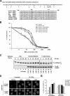
FIG. 1.
FIG. 1.
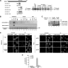
Two highly conserved Chk1 phosphorylation sites on FANCE. (A) Alignment of sequences surrounding the phosphorylation motif for Chk1 [R-X-X-(S/T)] in human FANCE with FANCE sequences in other organisms. (B) Complementation of MMC sensitivity of an FA-E lymphoblast cell line, EUFA130, with empty vector (pMMP), pMMP-FLAG-FANCE, pMMP-FLAG-T346A, pMMP-FLAG-S374A, and pMMP- FLAG-TS/AA. The indicated retroviral supernatants were generated and used to transduce EUFA130 cells. Puromycin-resistant cells were selected, and MMC sensitivity was determined as described in Materials and Methods. The values shown are the means ± standard deviations from four separate experiments. (C) Restoration of monoubiquitination of FANCD2. The indicated stably transduced FA-E lymphoblast cell lines were either untreated or exposed to IR at different doses, as indicated, and harvested after 6 h. Western blotting was performed with anti-FANCD2 or anti-FLAG antibodies. (D) Restoration of FANCD2 nuclear foci formation. EUFA130 lymphoblasts stably expressing FLAG-FANCEwt (EUFA130+FLAG-FANCE) and the double mutant FLAG-FANCE(TS/AA) (EUFA130+FLAG-TS/AA) were either untreated or treated with IR (10 Gy) and fixed 6 h later; immunofluorescence was determined using anti-FANCD2 (FI-17) antibody. Magnification, ×630. (E) Quantification of FANCD2 foci. Cells with more than four distinct foci were counted as positive; 200 cells/sample were analyzed. The values shown are the means ± standard deviations from three separate experiments. Vec, empty vector.
FIG. 2.
FIG. 2.
Phosphorylation of FANCE by Chk1 in vitro and in vivo. (A) GST-FANCE peptide fusion proteins, containing the indicated regions of FANCE, were generated and used as substrates for in vitro kinase assays; the threonine (346) and/or serine (374) residues that were mutated to alanine are shown (left panel). GST-FANCE peptide fusion proteins were incubated with [γ-32P]ATP and purified recombinant Chk1 as described in Materials and Methods. The reaction was stopped by the addition of SDS sample buffer before analysis by SDS-PAGE and autoradiography (right panel). The GST-Cdc25C(200-256) peptide fusion protein and GST were used as positive and negative control substrates, respectively, for Chk1 to demonstrate efficient in vitro kinase assays. (B) In vitro kinase assays using GST, purified recombinant Chk1, Chk2, or MAPKAPK2 (MK2) to phosphorylate the recombinant proteins (containing residues 149 to 536) rFANCEwt and the double mutant FANCE(rTS/AA) were performed and analyzed by SDS-PAGE, followed by immunoblotting with the pT346-FANCE and pS374-FANCE antibodies. The GST-Cdc25C(200-256) peptide fusion protein and GST were used as positive and negative control substrates, respectively, for Chk1, Chk2, and MK2 to demonstrate efficient in vitro kinase assays (data not shown). (C) Chk1 phosphorylates FANCE in vivo. EUFA130 lymphoblasts were stably expressed with empty vector (Vec), FLAG-FANCEwt, and FLAG-FANCE(TS/AA) as indicated. Cells were either untreated or treated with UV (60 J/m2); after 3 h, immunoprecipitation was performed using anti-FLAG antibody, followed by SDS-PAGE and then Western blotting with anti-pT346-FANCE and anti-pS374-FANCE phosphospecific antibodies and anti-FLAG antibody. (D) EUFA130 lymphoblasts stably expressing empty vector (EUFA130+Vec) and FLAG-FANCEwt (EUFA130+FLAG-FANCE) were either untreated or treated with UV (60 J/m2) and fixed 2 h later; immunofluorescence was performed using anti-pT346-FANCE antibody (left panel). Magnification, ×630. HeLa cells were transiently transfected with siRNA targeted against GFP (control), Chk1, or ATR. After 72 h of transfection, cells were either untreated or treated with UV (60 J/m2) and incubated for 2 h before fixation; immunofluorescence was performed using anti-pT346-FANCE antibody (right panel). Magnifi- cation, ×400. (E) Quantification of pT346-FANCE foci. Cells with more than four distinct foci were counted as positive; 100 cells/sample were analyzed. The values shown are the means ± standard deviations from three separate experiments. The formation of pT346 FANCE foci with the treatment of UV (60 J/m2) was strongly decreased in HeLa cells in which ATR or Chk1 had been suppressed with siRNA.
FIG. 3.
FIG. 3.
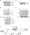
Knockdown of Chk1 results in an increased basal level of FANCD2 foci assembly. (A and B) HeLa cells were transfected with siRNAs targeted against GFP (control) or Chk1. After 72 h of transfection, cells were treated with UV (60 J/m2) and incubated for 3 h or 6 h before fixation and lysis; immunofluorescence was performed using anti-FANCD2 antibody. Magnification, ×400 (A) Whole-cell extracts were analyzed by Western blotting with the indicated antibodies. An anti-β-tubulin blot was used as a loading control (B). (C) Quantification of FANCD2 foci. Cells with more than four distinct foci were counted as positive; 200 cells/sample were analyzed. The values shown are the means ± standard deviations from three separate experiments.
FIG. 4.
FIG. 4.
Colocalization of pT346-FANCE foci and FANCD2-Ub foci after DNA damage. (A) The kinetics of pT346-FANCE foci and FANCD2 foci were followed after DNA damage. HeLa cells were either untreated or treated with UV (60 J/m2) and incubated for different periods of time (30 min, 2 h, 4 h, 6 h, or 8 h) as indicated before fixation; immunofluorescence was performed using anti-pT346-FANCE and anti-FANCD2 (FI-17) antibodies. Magnification, ×400. (B) Cells with more than four distinct foci were counted as positive; 200 cells/sample were analyzed. The values shown are the means ± standard deviations from three separate experiments. (C) After 4 h of UV irradiation, colocalization of pT346-FANCE foci and FANCD2 foci in HeLa cells is shown. Magnification, ×630.
FIG. 5.
FIG. 5.
FANCE phosphorylation by Chk1 does not correct MMC-mediated cell death but corrects cell cycle progression and promotes DNA synthesis following MMC treatment. (A) FA-E cells stably expressing wt FANCE (EUFA130+FLAG-FANCEwt) or the double mutant of FANCE (EUFA130+FLAG-TS/AA) do not accumulate in the late S/G2 phase of the cell cycle after 24 h of MMC treatment compared to cells stably expressing empty vector (EUFA130+Vec). The values shown are the means ± standard deviations from three separate experiments. (B) FA-E cells stably expressing empty vector (EUFA130+Vec) have decreased DNA synthesis after 24 h of MMC 160 ng/ml treatment compared to FA-E cells stably expressing wt FANCE (EUFA130+FLAG-FANCEwt) or the double mutant of FANCE (EUFA130+FLAG-TS/AA). The values shown are the means ± standard deviations from three separate experiments. (C) FA-E cells stably expressing the double mutant of FANCE (EUFA130+FLAG-TS/AA) or empty vector (EUFA130+Vec) demonstrate a higher percentage of sub-G1 cells after 48 and 72 h following MMC (160 ng/ml) treatment than cells expressing wt FANCE. Values shown are the means ± standard deviations from three separate experiments.
FIG. 6.
FIG. 6.
FANCE phosphorylation by Chk1 promotes its degradation. (A) HeLa cells were either untreated or treated with UV irradiation at 60 J/m2 and incubated for different periods of time as indicated before lysis. Whole-cell extracts were immunoblotted with the indicated antibodies. An anti-β-tubulin blot was used as a loading control. (B) HeLa cells were synchronized by a double-thymidine block and then released into S phase. One hour after release, the cells were either untreated (Control) or treated with UV (60 J/m2, 15 h), HU (2 mM, 24 h), MMC (160 ng/ml, 24 h), or cisplatin, (10 μM, 24 h). Whole-cell extracts were analyzed by Western blotting with the indicated antibodies. An anti-β-tubulin blot was used as a loading control. (C) EUFA130 lymphoblasts were stably expressed with pMMP (empty vector [Vec]), FLAG-FANCEwt, or FLAG-FANCE(TS/AA) (the double mutant) as indicated. Cells were either untreated or treated with UV (60 J/m2); after 8 h, whole-cell extracts were analyzed by Western blotting with the indicated antibodies. An anti-β-tubulin blot was used as a loading control. (D) U2OS cells were either untreated or treated with UV (60 J/m2) and incubated for 3 h with or without the addition of 25 μM MG132 to the indicated samples during the final 2 h in cell culture. Whole-cell extracts were analyzed by Western blotting with the indicated antibodies. An anti-β-tubulin blot was used as a loading control. (E) FANCE ubiquitination in vivo. U2OS stably expressing empty vector (a), FLAG-FANCEwt (b), or and FLAG-FANCE(TS/AA) (c) were transiently transfected without or with a cDNA encoding HA-Ub; after 48 h of transfection, cells were untreated or treated with UV (60 J/m2) and incubated for 2 h before cells were lysed in SDS denaturation buffer. FLAG-FANCEwt and the double mutant protein were isolated by anti-FLAG antibody immunoprecipitation. Immune complexes were run on SDS-PAGE gels and immunoblotted with anti-HA or anti-FLAG antibodies. (F) Schematic model showing the activation of the FA/BRCA pathway by the ATR-Chk1 pathway. DNA damage or replication arrest (MMC, UV, IR, or HU) activates the ATR-dependent phosphorylation of FANCD2 (6) and the Chk1-dependent phosphorylation of FANCE. Both monoubiquitinated FANCD2 and phosphorylated FANCE are required for MMC resistance. A nonubiquitinated mutant of FANCD2 (K561R) or the nonphosphorylated mutant FANCE(TS/AA) fails to correct MMC hypersensitivity.

Similar articles

Cited by

References

    1. Andreassen, P. R., A. D. D'Andrea, and T. Taniguchi. 2004. ATR couples FANCD2 monoubiquitination to the DNA-damage response. Genes Dev. 18:1958-1963. - PMC - PubMed
    1. Auerbach, A. D. 1993. Fanconi anemia diagnosis and the diepoxybutane (DEB) test. Exp. Hematol. 21:731-733. - PubMed
    1. de Winter, J. P., F. Leveille, C. G. van Berkel, M. A. Rooimans, L. van Der Weel, J. Steltenpool, I. Demuth, N. V. Morgan, N. Alon, L. Bosnoyan-Collins, J. Lightfoot, P. A. Leegwater, Q. Waisfisz, K. Komatsu, F. Arwert, J. C. Pronk, C. G. Mathew, M. Digweed, M. Buchwald, and H. Joenje. 2000. Isolation of a cDNA representing the Fanconi anemia complementation group E gene. Am. J. Hum. Genet. 67:1306-1308. - PMC - PubMed
    1. Dunn, J., M. Potter, A. Rees, and T. M. Runger. 2006. Activation of the Fanconi anemia/BRCA pathway and recombination repair in the cellular response to solar ultraviolet light. Cancer Res. 66:11140-11147. - PubMed
    1. Garcia-Higuera, I., T. Taniguchi, S. Ganesan, M. S. Meyn, C. Timmers, J. Hejna, M. Grompe, and A. D. D'Andrea. 2001. Interaction of the Fanconi anemia proteins and BRCA1 in a common pathway. Mol. Cell 7:249-262. - PubMed

Publication types

MeSH terms