

Invasion and persistence of Mycobacterium avium subsp. paratuberculosis during early stages of Johne's disease in calves
- PMID: 17296749
- PMCID: PMC1865790
- DOI: 10.1128/IAI.01739-06
Invasion and persistence of Mycobacterium avium subsp. paratuberculosis during early stages of Johne's disease in calves
Abstract
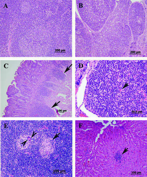
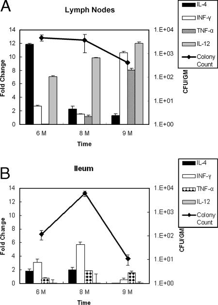
Infection with Mycobacterium avium subsp. paratuberculosis causes Johne's disease in cattle and is a serious problem for the dairy industry worldwide. Development of models to mimic aspects of Johne's disease remains an elusive goal because of the chronic nature of the disease. In this report, we describe a surgical approach employed to characterize the very early stages of infection of calves with M. avium subsp. paratuberculosis. To our surprise, strains of M. avium subsp. paratuberculosis were able to traverse the intestinal tissues within 1 h of infection in order to colonize distant organs, such as the liver and lymph nodes. Both the ileum and the mesenteric lymph nodes were persistently infected for months following intestinal deposition of M. avium subsp. paratuberculosis despite a lack of fecal shedding of mycobacteria. During the first 9 months of infection, humoral immune responses were not detected. Nonetheless, using flow cytometric analysis, we detected a significant change in the cells participating in the inflammatory responses of infected calves compared to cells in a control animal. Additionally, the levels of cytokines detected in both the ileum and the lymph nodes indicated that there were TH1-type-associated cellular responses but not TH2-type-associated humoral responses. Finally, surgical inoculation of a wild-type strain and a mutant M. avium subsp. paratuberculosis strain (with an inactivated gcpE gene) demonstrated the ability of the model which we developed to differentiate between the wild-type strain and a mutant strain of M. avium subsp. paratuberculosis deficient in tissue colonization and invasion. Overall, novel insights into the early stages of Johne's disease were obtained, and a practical model of mycobacterial invasiveness was developed. A similar approach can be used for other enteric bacteria.
Figures


Similar articles
-
Immune responses after oral inoculation of weanling bison or beef calves with a bison or cattle isolate of Mycobacterium avium subsp. paratuberculosis.J Wildl Dis. 2003 Jul;39(3):545-55. doi: 10.7589/0090-3558-39.3.545. J Wildl Dis. 2003. PMID: 14567215
-
New method of serological testing for Mycobacterium avium subsp. paratuberculosis (Johne's disease) by flow cytometry.Foodborne Pathog Dis. 2005 Fall;2(3):250-62. doi: 10.1089/fpd.2005.2.250. Foodborne Pathog Dis. 2005. PMID: 16156706
-
Cytokine gene expression in peripheral blood mononuclear cells and tissues of cattle infected with Mycobacterium avium subsp. paratuberculosis: evidence for an inherent proinflammatory gene expression pattern.Infect Immun. 2004 Mar;72(3):1409-22. doi: 10.1128/IAI.72.3.1409-1422.2004. Infect Immun. 2004. PMID: 14977946 Free PMC article.
-
Regulatory T cells in cattle and their potential role in bovine paratuberculosis.Comp Immunol Microbiol Infect Dis. 2012 May;35(3):233-9. doi: 10.1016/j.cimid.2012.01.004. Epub 2012 Jan 27. Comp Immunol Microbiol Infect Dis. 2012. PMID: 22285689 Review.
-
Current perspectives on Mycobacterium avium subsp. paratuberculosis, Johne's disease, and Crohn's disease: a review.Crit Rev Microbiol. 2011 May;37(2):141-56. doi: 10.3109/1040841X.2010.532480. Epub 2011 Jan 22. Crit Rev Microbiol. 2011. PMID: 21254832 Review.
Cited by
-
Evaluation of the immune status of peripheral blood monocytes from dairy cows during the periparturition period.J Reprod Dev. 2019 Aug 9;65(4):313-318. doi: 10.1262/jrd.2018-150. Epub 2019 May 2. J Reprod Dev. 2019. PMID: 31061297 Free PMC article.
-
Stereotypic immune response in Mycobacterium avium ssp. paratuberculosis infection among different Swiss caprine genotypes.Vet Pathol. 2025 Mar 17;62(5):3009858251322726. doi: 10.1177/03009858251322726. Online ahead of print. Vet Pathol. 2025. PMID: 40094295 Free PMC article.
-
Key role for the alternative sigma factor, SigH, in the intracellular life of Mycobacterium avium subsp. paratuberculosis during macrophage stress.Infect Immun. 2013 Jun;81(6):2242-57. doi: 10.1128/IAI.01273-12. Epub 2013 Apr 8. Infect Immun. 2013. PMID: 23569115 Free PMC article.
-
Divergent immune responses to Mycobacterium avium subsp. paratuberculosis infection correlate with kinome responses at the site of intestinal infection.Infect Immun. 2013 Aug;81(8):2861-72. doi: 10.1128/IAI.00339-13. Epub 2013 May 28. Infect Immun. 2013. PMID: 23716614 Free PMC article.
-
Cytokine and Chemokine Concentrations as Biomarkers of Feline Mycobacteriosis.Sci Rep. 2018 Nov 23;8(1):17314. doi: 10.1038/s41598-018-35571-5. Sci Rep. 2018. PMID: 30470763 Free PMC article.
References
-
- Ayele, W. Y., M. Bartos, P. Svastova, and I. Pavlik. 2004. Distribution of Mycobacterium avium subsp paratuberculosis in organs of naturally infected bull-calves and breeding bulls. Vet. Microbiol. 103:209-217. - PubMed
-
- Bannantine, J. P., J. F. J. Huntley, E. Miltner, J. R. Stabel, and L. E. Bermudez. 2003. The Mycobacterium avium subsp. paratuberculosis 35 kDa protein plays a role in invasin of bovine epithelial cells. Microbiology 149:2061-2069. - PubMed
-
- Bouma, G., and W. Strober. 2003. The immunological and genetic basis of inflammatory bowel disease. Nat. Rev. Immunol. 3:521-533. - PubMed
-
- Collins, M. T. 1996. Diagnosis of paratuberculosis. Vet. Clin. N. Am. Food Anim. Pract. 12:357-371. - PubMed
Publication types
MeSH terms
LinkOut - more resources
Full Text Sources
Molecular Biology Databases
