Comparative Study
. 2007 Feb 19;204(2):421-30.
doi: 10.1084/jem.20061604. Epub 2007 Feb 12.

Leukocyte-specific protein 1 interacts with DC-SIGN and mediates transport of HIV to the proteasome in dendritic cells

Affiliations
Comparative Study

Leukocyte-specific protein 1 interacts with DC-SIGN and mediates transport of HIV to the proteasome in dendritic cells

Alvin L Smith et al. J Exp Med. .

Abstract

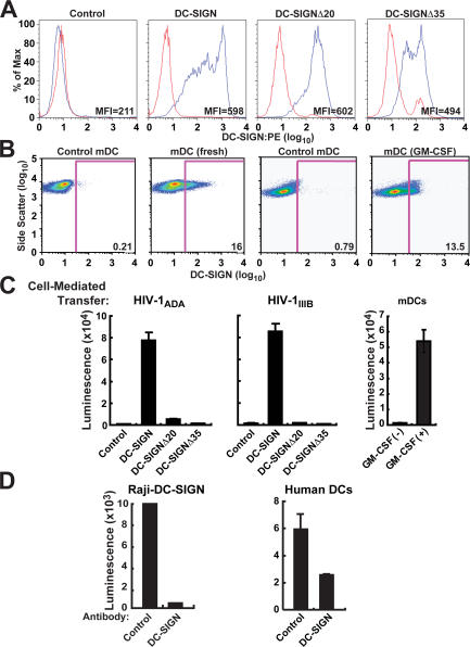
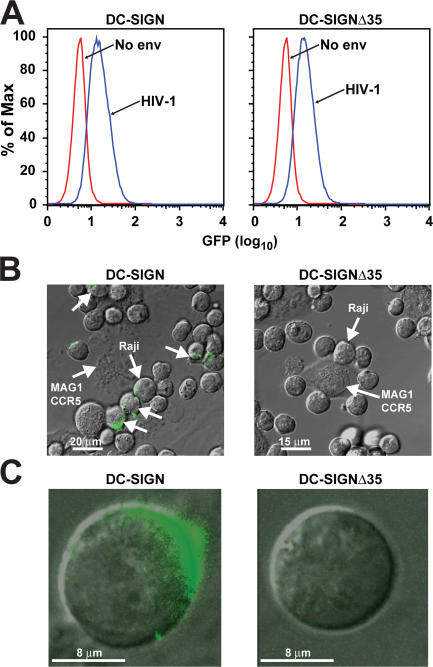
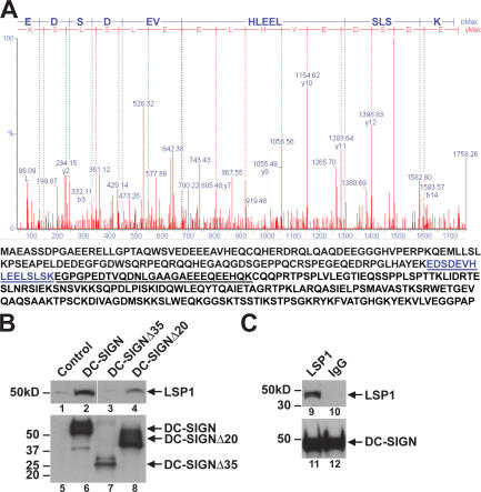
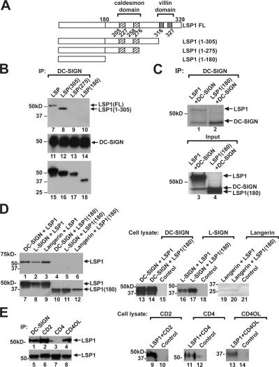
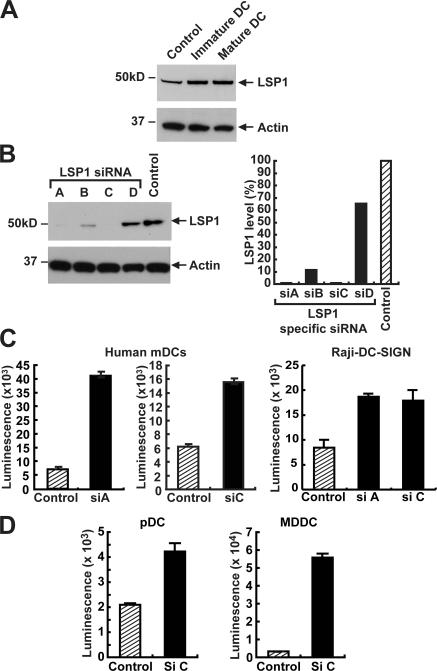
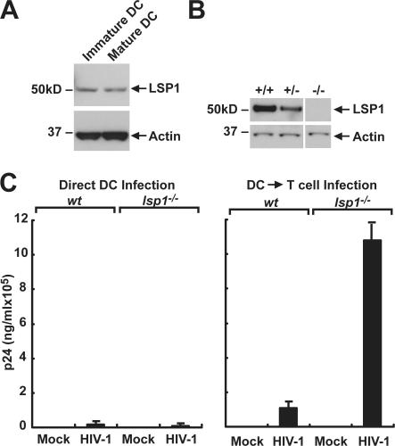
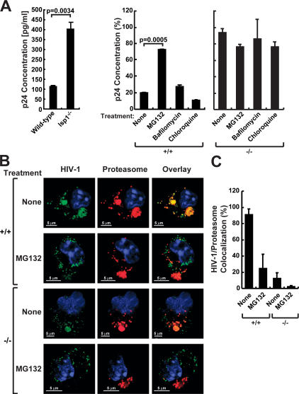
Dendritic cells (DCs) capture and internalize human immunodeficiency virus (HIV)-1 through C-type lectins, including DC-SIGN. These cells mediate efficient infection of T cells by concentrating the delivery of virus through the infectious synapse, a process dependent on the cytoplasmic domain of DC-SIGN. Here, we identify a cellular protein that binds specifically to the cytoplasmic region of DC-SIGN and directs internalized virus to the proteasome. This cellular protein, leukocyte-specific protein 1 (LSP1), was defined biochemically by immunoprecipitation and matrix-assisted laser desorption/ionization time-of-flight mass spectrometry. LSP1 is an F-actin binding protein involved in leukocyte motility and found on the cytoplasmic surface of the plasma membrane. LSP1 interacted specifically with DC-SIGN and other C-type lectins, but not the inactive mutant DC-SIGNDelta35, which lacks a cytoplasmic domain and shows altered virus transport in DCs. LSP1 diverts HIV-1 to the proteasome. Down-regulation of LSP1 with specific small interfering RNAs in human DCs enhanced HIV-1 transfer to T cells, and bone marrow DCs from lsp1(-/-) mice also showed an increase in transfer of HIV-1(BaL) to a human T cell line. Proteasome inhibitors increased retention of viral proteins in lsp1(+/+) DCs, and substantial colocalization of virus to the proteasome was observed in wild-type compared with LSP1-deficient cells. Collectively, these data suggest that LSP1 protein facilitates virus transport into the proteasome after its interaction with DC-SIGN through its interaction with cytoskeletal proteins.


Figures

Figure 1.
Figure 1.
DC-SIGN is necessary for HIV-1 uptake and transfer to T cells. (A) DC-SIGN expression on Raji B cells. Raji B cells expressing full-length DC-SIGN, DC-SIGNΔ35, and DC-SIGNΔ20 were incubated with human mAb DC-SIGN/PE for 30 min at 4°C, washed once with PBS, and analyzed by flow cytometry (MFI, mean fluorescence intensity). (B) DC-SIGN expression on human mDCs. Immature human mDCs were incubated with 2 μg of control or human mAb DC-SIGN (eBioscience) for 30 min at 4°C, washed once with PBS, and incubated with donkey anti–mouse Alexa 647–conjugated IgG for 30 min at 4°C, washed once with PBS, and analyzed by flow cytometry. (C) Uptake and transfer of CCR5- and CXCR4-tropic HIV-1 by DC-SIGN. Raji B cells expressing full-length DC-SIGN, DC-SIGNΔ35, and DC-SIGNΔ20 were pulsed with 750 ng/ml HIV-1ADA (CCR5-tropic) or HIV-1IIIB (CXCR4) at 37°C for 2 h, trypsinized, washed, and incubated with A3R5 or MT2 cells, respectively. Also, human mDCs were pulsed with ∼780 ng/ml HIV-1ADA at 37°C for 2 h, trypsinized, washed, and incubated with A3R5. All cells were lysed 72 h after incubation and analyzed for luciferase activity. (D) mAbs to DC-SIGN inhibit uptake and transfer of HIV-1 into T leukemia cells by Raji B cells or human DCs. Raji B cells (left) expressing full-length DC-SIGN and human DCs (right) were incubated with mAb to DC-SIGN for 30 min at 4°C before incubating with 750 ng/ml HIV-1ADA for 2 h at 37°C. Cells were then washed and incubated with MT2 T leukemia cells. Cells were lysed 72 h after incubation and analyzed for luciferase activity.
Figure 2.
Figure 2.
The cytoplasmic domain of DC-SIGN is necessary for HIV-1 internalization. (A) Raji B cells expressing full-length DC-SIGN and cytoplasmic 35 amino acid–deleted DC-SIGNΔ35 bind HIV-1. Approximately 780 ng/ml HIV-1 GFP-labeled (blue peak) and a No-Env negative control (red peak) virions were incubated with Raji cells expressing full-length and the cytoplasmic mutant DC-SIGNΔ35 for 30 min at 4°C. Cells were then analyzed by flow cytometry for cell surface–bound virus. (B and C) Raji cells expressing full-length DC-SIGN internalize HIV-1. Raji cells were incubated with HIV-1-GFP virions at 37°C for 2 h, trypsinized, washed, and added to HeLa cells expressing CD4/CCR5 (MAGI-CCR5) to visualize not only internalization, but polarization. Unlabeled arrows indicate polarization of virus between cells. Cells were viewed using confocal microscopy (B, low magnification; C, high magnification) and analyzed using Leica software.
Figure 3.
Figure 3.
LSP1 associates with the cytoplasmic domain of DC-SIGN. (A) Identification of proteins interacting with the cytoplasmic domain of DC-SIGN. Raji cells, expressing DC-SIGN, DC-SIGNΔ35, and DC-SIGNΔ20 were lysed in cell lysis buffer and 2 mg of total protein was incubated overnight with 5 μg of monoclonal anti–DC-SIGN antibody and protein G conjugated to agarose beads at 4°C. The immunoprecipitated material was washed and suspended in 2-D gel electrophoresis loading buffer. After 2-D gel electrophoresis, gels were stained with Coomassie blue. Coomassie-stained protein spots that were unique for the full-length DC-SIGN were picked, digested with trypsin, and analyzed by MALDI-TOF. A representation of the mass spectrometry data for the 15–amino acid LSP1 peptide is shown (top). Protein sequence of LSP1 is shown, and the peptides identified by matrix-assisted laser desorption/ionization time-of-flight are underlined (bottom). (B) LSP1 interacts with full-length DC-SIGN but not the cytoplasmic tail–deleted mutant. The parental cell line Raji-1 and Raji B cells expressing DC-SIGN and nonfunctional mutants DC-SIGNΔ35 and DC-SIGNΔ20 were lysed, and total protein was immunoprecipitated with monoclonal anti–DC-SIGN antibody and protein G conjugated to agarose beads overnight at 4°C. Immunoprecipitates were assayed for LSP1 (lanes 1–4) and DC-SIGN (lanes 5–8) expression using polyclonal antibodies by immunoblot. The control represents cell lysate from the parental cell line Raji-1. (C) LSP1 interacts with DC-SIGN in human DCs. Human MDDCs were isolated from healthy donors and cultured in RPMI, 50 ng/ml hGM-CSF, and 100 ng/ml IL-4 for 7 d before being lysed, and total protein was immunoprecipitated in the presence of protease inhibitors with a monoclonal anti–DC-SIGN antibody conjugated to agarose beads. Immunoprecipitates were assayed for LSP1 (lanes 9 and 10) and DC-SIGN (lanes 11 and 12) expression using polyclonal antibodies by immunoblot.
Figure 4.
Figure 4.
DC-SIGN associates with LSP1 through a region distinct from its actin binding domains. (A) The caldesmon-like CI and CII (hatched) and villin-like VI and VII (gray) on LSP1 that bind F-actin are indicated. The early truncation mutants generated by site-directed mutagenesis at amino acids 305, 275, and 180 are shown. (B) 293T cells (106 cells/well) in a six-well plate were cotransfected with full-length DC-SIGN and full-length LSP1 or the mutants (1–305, 275, and 180) as indicated. Cell lysates prepared 72 h after transfection were immunoprecipitated with anti–DC-SIGN antibody and assayed for LSP1 (lanes 7–10) and DC-SIGN (lanes 11–14) by immunoblot. Cell lysates of transfected cells were analyzed by immunoblot for LSP1 expression in full-length and mutant 1–305, 1–275, and 1–180 constructs (lanes 15–18). (C) 35S-labeled in vitro–cotranslated full-length LSP1 interacts directly with DC-SIGN. Full-length LSP1 or LSP1(1–180) cDNA were in vitro cotranslated with DC-SIGN in rabbit reticulocytes in the presence of 35S. Labeled proteins were immunoprecipitated in the presence of protease inhibitors with monoclonal anti–DC-SIGN conjugated to agarose beads, electrophoresed on a 10% polyacrylamide gel, and visualized by autoradiography. LSP1 and DC-SIGN (lanes 1 and 3), DC-SIGN (lanes 2 and 4), and LSP1(1–180) (lane 4) are shown. (D) 293T cells (106 cells/well) in a six-well plate were cotransfected with full-length LSP1 or LSP1(1–180) along with DC-SIGN, L-SIGN, and Langerin. Cell lysates prepared 72 h after transfection were immunoprecipitated with anti–DC-SIGN, L-SIGN, or Langerin or mAbs, and immunoblotted with polyclonal antibody to LSP1 (lanes 1–6). Expression of full-length LSP1 and LSP1(1–180) in the cell lysate (lanes 7–12) is shown. DC-SIGN (lanes 13 and 14), L-SIGN (lanes 16 and 17), and Langerin (lanes 19 and 20) expression in the cell lysates was assayed by immunoblot. The control is lysate from 293T cells transfected with a GFP plasmid. (E) LSP1 interacts with other extracellular surface molecules. 293T cells (106 cells/well) in a six-well plate were cotransfected with full-length LSP1 along with DC-SIGN, CD2, CD4, and CD40L. Cell lysates prepared 72 h after transfection were immunoprecipitated with anti–DC-SIGN, CD2, CD4, and CD40L mAbs and immunoblotted with polyclonal antibody to LSP1 (lanes 1–4), or expression of full-length LSP1 in the cell lysate was determined (lanes 5–8). CD2 (lanes 9 and 10), CD4 (lanes 11 and 12), and CD40L (lanes 13 and 14) expression in the cell lysates were assayed by immunoblotting. The control is lysate from 293T cells transfected with a GFP plasmid.
Figure 5.
Figure 5.
LSP1 down-regulation causes increased HIV-1 transfer in human DCs. (A) LSP1 expression in human DCs. Cell lysates prepared from immature or poly:IC-matured mDCs isolated from human elutriated monocytes were assayed for LSP1 expression by a polyclonal anti-LSP1 antibody. Expression was measured against control cell lysates from 293T transfected with the LSP1 gene. (B) Specific siRNAs for human LSP1 cause a down-regulation of LSP1 expression. Raji–DC-SIGN cells were transfected with the indicated LSP1, and control siRNA duplexes were introduced by nucleofection. 24 h after transfection, cell lysates (20 μg of total protein) were assayed for LSP1 expression by immunoblot (left). The film images were digitized using an Epson scanner and quantified using Bio-Rad Quantity One software. The numbers obtained for LSP1 were corrected using the numbers obtained from actin and plotted relative to control in a Microsoft Excel graph as indicated (right). (C and D) Down-regulation of human LSP1 in human DCs and Raji DC-SIGN cell line causes an increase in HIV-1 transfer to T cells. Mature human DCs were transiently transfected by GeneSilencer with siRNA for LSP1 (siA and siC) or scrambled siRNA (control). To label transfected cells, unrelated Cy5 siRNA was mixed with both control siRNA and LSP1 siRNAs at a ratio of 4:1. 8 h after the siRNA transfection, the cells were sorted for the Cy5 label and plated onto 96-well plates. 24 h after transfection, cells were pulsed with luciferase expressing 780 ng/ml HIV-1ADA for 2 h at 37°C, washed five times, and incubated with A3R5 T cells. Cells were lysed 72 h after transfection and analyzed for luciferase activity. These experiments were performed in three independent HIV-1 donors in triplicate samples, and two such experiments are represented in the figure.
Figure 6.
Figure 6.
Murine DCs from lsp1−/− mice increase HIV-1 uptake and transfer to T cells. (A) LSP1 expression in murine DCs remains constant after maturation. Cell lysates prepared from immature or CpG-matured bone marrow–derived murine DCs were assayed for LSP1 expression by a polyclonal anti-LSP1 antibody. (B) LSP1 expression in mature BMDCs isolated from lsp1−/−, lsp1+/−, and lsp1+/+ littermates. Cell lysates prepared from CpG-matured bone marrow–derived murine DCs were assayed for LSP1 expression by a polyclonal anti-LSP1 antibody. (C) LSP1-null BMDCs enhance HIV-1 trans-infection of T cells. Mature BMDCs (105/well) from WT (wt) or lsp1−/− mice were pulsed with HIV-1BaL (∼780 ng/ml of p24/ml) or RPMI alone for 2 h at 37°C. Cells were washed extensively to remove free virus and replaced with fresh media, and A3R5 T cells were added to one set of mock or HIV-1–infected BMDCs followed by incubation for another 72 h. At the appropriate time, cell supernatants were collected, and p24 ELISA was performed as instructed by the manufacturer (Coulter). The data are representative of duplicate experiments.
Figure 7.
Figure 7.
LSP1 traffics HIV-1 to the proteasome. (A) HIV-1 degradation is decreased in lsp1−/− DCs, and proteasome inhibitors block HIV-1 degradation in the presence of LSP1. Mature BMDCs from lsp1−/− and wt mice were pulsed with HIV-1–GFP for 2 h at 37°C, washed with PBS, trypsinized, and lysed with p24 lysis buffer reagent (left). Mature BMDCs from wt (lsp1+/+) were pretreated for 1 h with MG132 (proteasome inhibitor), chloroquine, or bafilomycin A-1 (lysosomal inhibitors) in RPMI or RPMI alone in a 96-well tissue culture dish (5 × 10⁴ cells). Cells were then pulsed with HIV-1–GFP for 2 h at 37°C in the presence of inhibitors, washed with PBS, replaced with media containing the respective inhibitors, and incubated for an additional 1 h. Cells were then trypsinized, washed, and lysed with p24 lysis buffer reagent (right). p24 Gag concentrations were assayed by ELISA. These data are representative of the 1-h time point from multiple experiments performed in triplicate. The percentage values were calculated by setting the amount of p24 measured immediately after removing the virus from the cells, the 0-h time point, to 100%. The differences in p24 after several hours were then measured and compared with the 0-h time point. (B and C) HIV-1 shows greater colocalization with the proteasome in DCs expressing LSP1. (B) Mature BMDCs from lsp1−/− (−/−) and wt (+/+) mice were pulsed with HIV-1–GFP (green) for 30 min at 37°C. For proteasome inhibitor studies, BMDCs from lsp1−/− and wt mice were incubated with the proteasomal inhibitor MG132 in 5 μg/ml RPMI or RPMI alone for 1 h before infection by HIV-1–GFP (green) for 30 min. Cells were washed, fixed, and stained with a mAb to the 20S proteasome subunit a-4 (red) and viewed using confocal microscopy. HIV-1 that colocalizes with proteasomes appears yellow (overlay). (C) HIV-1 colocalization with proteasomes is representative of a percentage of HIV-1 that colocalizes with a percentage of proteasomes above a measurable threshold. Percentage colocalization was calculated using the Leica confocal software. Three images/sample were used, and error bars were assigned accordingly.

Similar articles

Cited by

References

    1. Steinman, R.M., and K. Inaba. 1999. Myeloid dendritic cells. J. Leukoc. Biol. 66:205–208. - PubMed
    1. Mellman, I., and R.M. Steinman. 2001. Dendritic cells: specialized and regulated antigen processing machines. Cell. 106:255–258. - PubMed
    1. Steinman, R.M., and M.C. Nussenzweig. 2002. Avoiding horror autotoxicus: the importance of dendritic cells in peripheral T cell tolerance. Proc. Natl. Acad. Sci. USA. 99:351–358. - PMC - PubMed
    1. Steinman, R.M. 1991. The dendritic cell system and its role in immunogenicity. Annu. Rev. Immunol. 9:271–296. - PubMed
    1. Chesnut, R.W., and H.M. Grey. 1985. Antigen presenting cells and mechanisms of antigen presentation. Crit. Rev. Immunol. 5:263–316. - PubMed

Publication types

MeSH terms