. 2007 Feb 12;176(4):535-44.
doi: 10.1083/jcb.200611058.

Hyaluronan and CD44 antagonize mitogen-dependent cyclin D1 expression in mesenchymal cells

Affiliations

Hyaluronan and CD44 antagonize mitogen-dependent cyclin D1 expression in mesenchymal cells

Devashish Kothapalli et al. J Cell Biol. .

Abstract

High molecular weight (HMW) hyaluronan (HA) is widely distributed in the extracellular matrix, but its biological activities remain incompletely understood. We previously reported that HMW-HA binding to CD44 antagonizes mitogen-induced S-phase entry in vascular smooth muscle cells (SMCs; Cuff, C.A., D. Kothapalli, I. Azonobi, S. Chun, Y. Zhang, R. Belkin, C. Yeh, A. Secreto, R.K. Assoian, D.J. Rader, and E. Puré. 2001. J. Clin. Invest. 108:1031-1040); we now characterize the underlying molecular mechanism and document its relevance in vivo. HMW-HA inhibits the mitogen-dependent induction of cyclin D1 and down-regulation of p27(kip1) in vascular SMCs. p27(kip1) messenger RNA levels were unaffected by HMW-HA, but the expression of Skp2, the rate-limiting component of the SCF complex that degrades p27(kip1), was reduced. Rescue experiments identified cyclin D1 as the primary target of HMW-HA. Similar results were observed in fibroblasts, and these antimitogenic effects were not detected in CD44-null cells. Analysis of arteries from wild-type and CD44-null mice showed that the effects of HMW-HA/CD44 on cyclin D1 and Skp2 gene expression are detected in vivo and are associated with altered SMC proliferation after vascular injury.


Figures
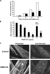
Figure 1.
Figure 1.
HMW-HA inhibits S-phase entry in human SMCs. (A) Quiescent human vascular SMCs were stimulated with 10% FBS for the selected times in the absence (control; white bars) or presence (black bars) of 200 μg/ml HMW-HA. *, P < 0.001. (B) Quiescent human vascular SMCs were stimulated with 10% FBS for 48 h in the presence of increasing concentrations of HMW-HA. A blocking antibody specific for human CD44 (5F12) or 50 μg/ml of an isotype-matched irrelevant antibody was added to the cells at the time of FBS stimulation and remained in the culture for the duration of the experiment. BrdU incorporation into nuclei was determined by immunofluorescence microscopy. *, P < 0.01; **, P < 0.001. (C) Quiescent human SMCs were stimulated with 10% FBS in the presence or absence of 200 μg/ml HMW-HA for 24 h. The cells were then stained with fluorescein-phalloidin and antivinculin. FBS-stimulated BrdU incorporation was inhibited 54% by HMW-HA in this experiment. Bars, 20 μm.
Figure 2.
Figure 2.
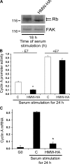
Pocket protein–dependent effects of HMW-HA on cyclin A gene transcription. Quiescent human vascular SMCs (G0) were stimulated with 10% FBS for the selected times in the absence (control, C) or presence of 200 μg/ml HMW-HA. (A) The cells were collected, lysed, and immunoblotted for Rb and focal adhesion kinase (FAK; loading control). Top and bottom arrows indicate the positions of hyper- and hypophosphorylated Rb, respectively. Quantification of Rb gel shifts from four independent experiments indicated that Rb phosphorylation after FBS stimulation (18–24 h) was 54 and 28% in the absence and presence of HMW-HA, respectively. (B) Human SMCs were cotransfected with plasmids encoding the cyclin A promoter-driving luciferase, empty vector or E7, and Renilla luciferase. The transfected cells were serum starved for 24 h and stimulated with 10% FBS. Cyclin A promoter activity was determined in the absence or presence of HMW-HA and E7. Cyclin A promoter-luciferase activity is normalized to Renilla luciferase activity. *, P < 0.01. (C) Quiescent (G0) human SMCs were stimulated with 10% FBS for 24 h. Total RNA was isolated and analyzed by QPCR for cyclin A mRNA and 18S rRNA. Cyclin A mRNA expression is normalized to 18S rRNA. *, P < 0.001. Error bars represent SD.
Figure 3.
Figure 3.
Subcellular effects of HMW-HA on G1-phase cdks. Quiescent human vascular SMCs (G0) were stimulated with 10% FBS for the selected times in the absence (control, C) or presence of 200 μg/ml HMW-HA. (A) Collected cells were lysed and immunoblotted for cyclin D1, p27, p21, cyclin A, and cdk4 (loading control). (B and C) Total RNA was isolated from SMCs and analyzed by QPCR for cyclin D1 mRNA, p27 mRNA, and 18S rRNA, and the levels of cyclin D1 (B) and p27 mRNA (C) were normalized to 18S rRNA. *, P < 0.01. (D) Quiescent human SMCs were stimulated with 10% FBS for 24 h in the absence or presence of 200 μg/ml HMW-HA. Total RNA was isolated from the SMCs and analyzed by QPCR for Skp2 mRNA and 18S rRNA. Skp2 mRNA expression is plotted relative to 18S rRNA. *, P < 0.001. Collected cells were also lysed and immunoblotted for Skp2 and cdk4 (inset). Error bars represent SD.
Figure 4.
Figure 4.
Cyclin D1 is the proximal target of HMW-HA. Human vascular SMCs infected with adenoviruses encoding lacZ (Ad-lacZ), cyclin D1 (Ad-D1), or Skp2 (Ad-Skp2) were serum starved and stimulated with 10% FBS-DME in the absence (control, C) or presence of 200 μg/ml HMW-HA. (A) The cultures were incubated for 24 h in the presence of BrdU, and BrdU incorporation into nuclei was determined by immunofluorescence microscopy. *, P < 0.01. (B) Collected cells were lysed and immunoblotted for cyclin D1 and cdk4 (loading control) or Skp2 and focal adhesion kinase (FAK; loading control). The thin vertical spaces between the Ad-lacZ, Ad-D1, and Ad-Skp2 images indicate reordering of these image blocks from the scanned gel. (C and D) Total RNA was isolated from the SMCs and used to determine the levels of Skp2 mRNA or cyclin D1 mRNA. The expression of cyclin D1 and Skp2 mRNAs was normalized to 18S rRNA and plotted relative to their expression levels in cells infected with the lacZ adenovirus. *, P < 0.005. Error bars represent SD.
Figure 5.
Figure 5.
HMW-HA inhibits cyclin D1, Skp2, and cyclin A expression in wild-type mouse vascular SMCs. Quiescent (G0) mouse vascular SMCs from wild-type (WT) or CD44-null mice were stimulated with 10% FBS for the selected time intervals in the absence (control, C) or presence of 200 μg/ml HMW-HA. (A) Cells were incubated for 24 h in the presence of BrdU, and BrdU incorporation into nuclei was determined by immunofluorescence microscopy. *, P < 0.002. (B) Cells were collected, lysed, and analyzed by QPCR for cyclin D1, Skp2, cyclin A, and p27 mRNAs as well as for 18S rRNA. Results for each mRNA are plotted relative to 18S rRNA. *, P < 0.01. (C) Collected cells from identical experiments were analyzed by Western blotting for either cyclin D1, Skp2, and cdk4 (loading control) or p27 and cdk4 (loading control). (D) Primary MEFs and vascular SMCs were lysed and immunoblotted for CD44. HA binding to CD44 was determined using FITC-conjugated HA in the absence and presence of CD44-neutralizing antibody (KM81). Error bars represent SD.
Figure 6.
Figure 6.
CD44 regulates the expression of cyclin D1, Skp2, and cyclin A in the aorta. (A and B) Total RNA was isolated twice from pooled, cleaned aortae of four 20-wk-old male CD44 wild-type (black bars) and CD44-null (white bars) mice for a total of eight mice/genotype. cDNA was synthesized for each isolation, and QPCR was performed on a cDNA aliquot for cyclin D1, Skp2, cyclin A mRNAs, and 18S rRNA (A) or CD31, CD68, smooth muscle actin (SMA) mRNAs, and 18S rRNA (B). The expression of cyclin D1, Skp2, cyclin A, CD31, CD68, and smooth muscle actin mRNAs are plotted relative to 18S rRNA. *, P < 0.003. Error bars represent SD. (C) Thoracic aortae were isolated from 20-wk-old male wild-type (WT) and CD44-null mice. The intima and adventitia were not removed before the tissues were fixed, embedded in paraffin, sliced in cross section, and stained with hematoxylin and eosin. Genotypes were confirmed by PCR. A, adventitia; M, media; L, lumen. Bars, 210 μm.
Figure 7.
Figure 7.
Increased neointima formation and BrdU incorporation after vascular injury in CD44-null mice. (A) Injured femoral arteries from wild-type and CD44-null mice were perfusion fixed, paraffin embedded, and sectioned before being stained for elastin. Boxes indicate the regions of the aortae that are shown at twofold higher magnification. Arrows indicate sites of neointima formation. (B) Intimal/medial ratios and the number of BrdU-positive nuclei in injured femoral arteries of wild-type and CD44-null mice. n = 5 per genotype. P = 0.02 for intimal/medial ratios, and P = 0.06 for BrdU as determined using a one-tailed t test. Horizontal bars indicate mean values. (C) The sham-operated control arteries from the mice in A were stained for elastin.

Similar articles

Cited by

References

    1. Aruffo, A., I. Stamenkovic, M. Melnick, C.B. Underhill, and B. Seed. 1990. CD44 is the principal cell surface receptor for hyaluronate. Cell. 61:1303–1313. - PubMed
    1. Bagui, T.K., S. Mohapatra, E. Haura, and W.J. Pledger. 2003. P27Kip1 and p21Cip1 are not required for the formation of active D cyclin-cdk4 complexes. Mol. Cell. Biol. 23:7285–7290. - PMC - PubMed
    1. Banerji, S., J. Ni, S.X. Wang, S. Clasper, J. Su, R. Tammi, M. Jones, and D.G. Jackson. 1999. LYVE-1, a new homologue of the CD44 glycoprotein, is a lymph-specific receptor for hyaluronan. J. Cell Biol. 144:789–801. - PMC - PubMed
    1. Bono, P., K. Rubin, J.M. Higgins, and R.O. Hynes. 2001. Layilin, a novel integral membrane protein, is a hyaluronan receptor. Mol. Biol. Cell. 12:891–900. - PMC - PubMed
    1. Bradshaw, A.D., and E.H. Sage. 2001. SPARC, a matricellular protein that functions in cellular differentiation and tissue response to injury. J. Clin. Invest. 107:1049–1054. - PMC - PubMed

Publication types

MeSH terms