Case Reports
. 2007 Feb;125(2):259-67.
doi: 10.1001/archopht.125.2.259.

Novel phenotypic and genotypic findings in X-linked retinoschisis

Affiliations
Case Reports

Novel phenotypic and genotypic findings in X-linked retinoschisis

Stephen H Tsang et al. Arch Ophthalmol. 2007 Feb.

Abstract

Objective: To describe atypical phenotypes associated with the retinoschisis (X-linked, juvenile) 1 mutation (RS1).

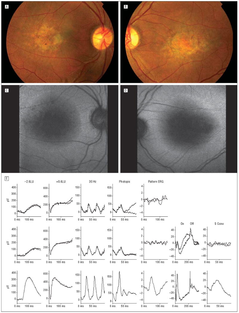
Methods: Seven patients with multiple fine white dots at the macula and reduced visual acuity were evaluated. Six patients underwent pattern and full-field electroretinography (ERG). On-off ERG, optical coherence tomography, and fundus autofluorescence imaging were performed in some patients. Mutational screening of RS1 was prompted by the ERG findings.

Results: Fine white dots resembling drusenlike deposits and sometimes associated with retinal pigment epithelial abnormalities were present in the maculae. An electronegative bright-flash ERG configuration was present in all patients tested, and abnormal pattern ERG findings confirmed macular dysfunction. A parafoveal ring of high-density autofluorescence was present in 3 eyes; 1 patient showed high-density foci concordant with the white dots. Optical coherence tomography did not show foveal schisis in 3 of 4 eyes. All patients carried mutations in RS1, including 1 with a novel 206T-->C mutation in exon 4.

Conclusions: Multiple fine white dots at the macula may be the initial fundus feature in RS1 mutation. Electrophysiologic findings suggest dysfunction after phototransduction and enable focused mutational screening. Autofluorescence imaging results suggest early retinal pigment epithelium involvement; a parafoveal ring of high-density autofluorescence has not previously been described in this disorder.


Figures

Figure 1
Figure 1
Various imaging findings in patient 1. A, and B, Fundus photographs showed fine intraretinal white lesions along the venules and arterioles of the perifoveolar vascular network. Retinal pigment epithelium disturbance associated with mildly thickened retina is seen over the superior part of the macula of the left eye. C, Red-free photograph of the right eye. D, Red-free photograph of the left eye. E and F, Confocal scanning laser ophthalmoscopic images revealed increased autofluorescence signal associated with the white dots. G, Foveal schisis was not detected by means of optical coherence tomography in the right macula. H, Parafoveal and peripheral microcystic changes were seen via optical coherence tomography in the left eye. I, Full-field and pattern electroretinograms (ERGs). Rows 1 and 2 show recordings from the right and left eyes, respectively, of the patient; row 3, normal recordings. LU indicates log units. Ordinal axes are shown in microvolts, allowing measurement of ERG amplitudes.
Figure 2
Figure 2
Fundus photographs (A and B) and fundus autofluorescence (C and D) in patient 2. Fine intraretinal white dots were seen in and adjacent to the central atrophic retinal pigment epithelium. Autofluorescence showed low-density signal over macular atrophy surrounded by a ring of high density. The observed ring of hyperfluorescence was consistent with retinal pigment epithelium disturbance outside of atrophic areas. E, Full-field and pattern electroretinograms (ERGs). Rows 1 and 2 show recordings from the right and left eyes, respectively, of the patient; row 3, normal recordings. LU indicates log units. Ordinal axes are shown in microvolts, allowing measurement of ERG amplitudes.
Figure 3
Figure 3
Fundus photographs (A and B) and autofluorescence (C and D) in patient 3 showing multifocal fine intraretinal white dots in the foveal regions. Low-density autofluorescence at the maculae, larger in the right eye (C), was surrounded by a ring of high density, suggesting a retinal pigment epithelium disturbance outside of atrophic areas. E, Full-field and pattern electroretinograms (ERGs). Rows 1 and 2 show recordings from the right and left eyes, respectively, of the patient; row 3, normal recordings. LU indicates log units. Ordinal axes are shown in microvolts, allowing measurement of ERG amplitudes.
Figure 4
Figure 4
Fundus photographs in patients 4 (A and B), 5 (C and D), 6 (E and F), and 7 (G and H) showing numerous white dots at the macula in all cases.

References

    1. Deutman AF, Pickers AJL, Aan de Kerk AL. Dominantly inherited cystoid macular edema. Am J Ophthalmol. 1976;82:540–548. - PubMed
    1. Traboulsi E, editor. Genetic Disease of the Eye. Oxford, England: Oxford University Press; 1998. Oxford Monographs on Medical Genetics; No. 36.
    1. George ND, Yates JR, Moore AT. X linked retinoschisis. Br J Ophthalmol. 1995;79:697–702. - PMC - PubMed
    1. Pimenides D, George ND, Yates JR, et al. X-linked retinoschisis: clinical phenotype and RS1 genotype in 86 UK patients. J Med Genet. 2005;42:e35. - PMC - PubMed
    1. Kellner U, Brummer S, Foerster MH, Wessing A. X-linked congenital retinoschisis. Graefes Arch Clin Exp Ophthalmol. 1990;228:432–437. - PubMed

Publication types