. 2007 Mar;129(3):221-31.
doi: 10.1085/jgp.200609658. Epub 2007 Feb 12.

Corelease and differential exit via the fusion pore of GABA, serotonin, and ATP from LDCV in rat pancreatic beta cells

Affiliations

Corelease and differential exit via the fusion pore of GABA, serotonin, and ATP from LDCV in rat pancreatic beta cells

Matthias Braun et al. J Gen Physiol. 2007 Mar.

Abstract

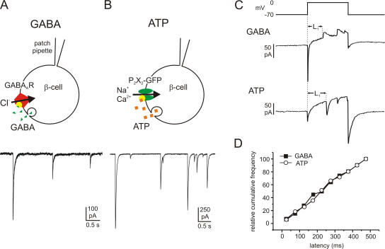
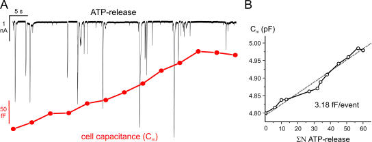
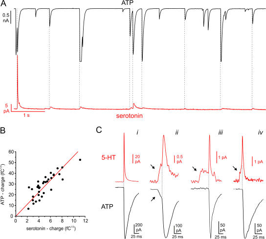
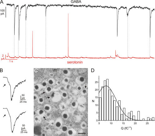
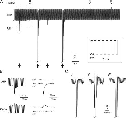
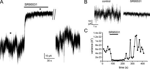
The release of gamma-aminobutyric acid (GABA) and ATP from rat beta cells was monitored using an electrophysiological assay based on overexpression GABA(A) or P2X2 receptor ion channels. Exocytosis of LDCVs, detected by carbon fiber amperometry of serotonin, correlated strongly (approximately 80%) with ATP release. The increase in membrane capacitance per ATP release event was 3.4 fF, close to the expected capacitance of an individual LDCV with a diameter of 0.3 microm. ATP and GABA were coreleased with serotonin with the same probability. Immunogold electron microscopy revealed that approximately 15% of the LDCVs contain GABA. Prespike "pedestals," reflecting exit of granule constituents via the fusion pore, were less frequently observed for ATP than for serotonin or GABA and the relative amplitude (amplitude of foot compared to spike) was smaller: in some cases the ATP-dependent pedestal was missing entirely. An inward tonic current, not dependent on glucose and inhibited by the GABA(A) receptor antagonist SR95531, was observed in beta cells in clusters of islet cells. Noise analysis indicated that it was due to the activity of individual channels with a conductance of 30 pS, the same as expected for individual GABA(A) Cl- channels with the ionic gradients used. We conclude that (a) LDCVs accumulate ATP and serotonin; (b) regulated release of GABA can be accounted for by exocytosis of a subset of insulin-containing LDCVs; (c) the fusion pore of LDCVs exhibits selectivity and compounds are differentially released depending on their chemical properties (including size); and (d) a glucose-independent nonvesicular form of GABA release exists in beta cells.


Figures

Figure 1.
Figure 1.
Quantal GABA and ATP release in rat β cells monitored by overexpression of the ionotropic membrane receptors GABAA and P2X2. Examples of GABA- (A) and ATP-activated (B) TICs recorded from β cells infected with GABAA and P2X2 receptors, respectively. Cells were held at −70 mV and exocytosis elicited by intracellular application of 2 μM free Ca2+. The cartoons above the current traces illustrate schematically the protocols used. (C) Samples of GABA (transient outward currents; top trace) and ATP release events (transient inward currents; bottom trace) triggered by 500-ms depolarizations from −70 to 0 mV. The latency between the beginning of the depolarizing pulse and the onset of the first exocytotic event during the pulse (L1) was measured. (D) Cumulative frequency of the first latencies of GABA release (black squares) and ATP release (open circles).
Figure 2.
Figure 2.
Estimation of the unitary LDCV capacitance increase. (A) P2X2 receptor–mediated TICs (top) and increase in whole-cell capacitance (bottom) recorded simultaneously in the same cell. Exocytosis was elicited by inclusion of 230 nM free Ca2+ in the pipette solution. Each segment (5 s) was preceded by a brief prepulse with a sine wave to measure the cell capacitance. (B) Plot of the cumulative number (ΣN) of purinergic TICs vs. membrane capacitance (Cm) from the same experiment. A linear fit (dotted line) with a slope of 3.18 fF/event is superimposed.
Figure 3.
Figure 3.
Parallel recordings of exocytotic events combining amperometry with overexpression of P2X2 receptors. The cells were held at −70 mV and exocytosis elicited by intracellular application of 2 μM free Ca2+. (A) Parallel recording of ATP release–induced TICs (top) and serotonin release measured by amperometry (bottom) in a single β-cell. The dotted lines indicate the simultaneous occurrence of TICs and amperometric spikes. (B) Cubic root of the charge of the P2X2-mediated TICs (n = 34 events recorded in the same cell) plotted against the cubic root of the charge of simultaneously recorded amperometric spikes. A linear fit is superimposed. (C) Examples of amperometric spikes (top red traces, 5-HT) and simultaneously recorded ATP release–induced TICs (bottom black trace). Prespike feet are indicated by arrows.
Figure 4.
Figure 4.
Corelease of GABA and serotonin by exocytosis of LDCVs. (A) Parallel recording of GABA-induced TICs (top) and serotonin release (bottom) in a single β-cell. The cell was held at −70 mV and exocytosis elicited by intracellular application of 2 μM free Ca2+. (B) Examples of GABA-induced TICs displaying prespike feet. (C) Immunogold labeling of GABA in rat pancreatic β cells. Labeling over the insulin-containing LDCVs (arrows) was less than that in the cytoplasm and was variable in density. (m = mitochondria; Bar, 200 nm). (D) Distribution of cubic roots of integrated GABA-dependent TICs (3√Q) in a single representative experiment. A total of 200 events (N) were analyzed. A Gaussian fit is superimposed. The CV in this particular experiment was 0.49.
Figure 5.
Figure 5.
Parallel monitoring of GABA and ATP release. (A, inset) Schematic of voltage protocol used to near-simultaneously resolve GABAergic and purinergic transient currents in cells infected with both GABAA and P2X2 receptors. The membrane potential was rapidly (50 Hz) alternated between ECl (−60 mV) and EP2X2 (+10 mV). (A) Sample recording showing ATP-induced inward currents (black arrows) and GABA-induced outward currents (white arrows) in a double-infected β-cell. Exocytosis was triggered by infusion of 0.2 μM free Ca2+ through the recording electrode. The largest inward currents have been truncated for display purposes. (B) Examples of an ATP-induced inward current and a GABA-induced outward response, marked by dashed boxes in A, shown on an expanded time base. In the right part, the current artifacts when switching between −60 and +10 mV have been removed and only current components at +10 and −60 mV are shown. The segments of the current recordings at the respective voltages have been connected by gray lines. (C) Examples of different types of events with a dominant inward current component: (i) inward current in isolation; (ii) clear out- and inward components; (iii) an outward component superseded by the activation of the inward current. The inward currents at −60 mV have been truncated for display purposes.
Figure 6.
Figure 6.
Evidence for tonic GABA release from β cells. (A) The recording was made in a β-cell overexpressing GABAA receptor Cl channels within a cell cluster. The holding current at −70 mV was measured in the presence of 1 mM extracellular glucose with intracellular solution containing 10 mM EGTA. The GABAA receptor antagonist SR95531 (10 μM) was added to the bath as indicated by the bar. A segment of ∼2 min during the washout of SR95531 has been removed. (B) Current segments marked by asterisks (*) in A shown at expanded time base. (C) Variance of current before, during, and after addition of SR95531 (as indicated by the horizontal line).

Similar articles

Cited by

References

    1. Ämmälä, C., F.M. Ashcroft, and P. Rorsman. 1993. Calcium-independent potentiation of insulin release by cyclic AMP in single β cells. Nature. 363:356–358. - PubMed
    1. Aspinwall, C.A., L. Huang, J.R. Lakey, and R.T. Kennedy. 1999. Comparison of amperometric methods for detection of exocytosis from single pancreatic β cells of different species. Anal. Chem. 71:5551–5556. - PubMed
    1. Barg, S. 2003. Mechanisms of exocytosis in insulin-secreting B-cells and glucagon-secreting A-cells. Pharmacol. Toxicol. 92:3–13. - PubMed
    1. Birnir, B., M.L. Tierney, N.P. Pillai, G.B. Cox, and P.W. Gage. 1995. Rapid desensitization of α1 β1 GABAA receptors expressed in Sf9 cells under optimized conditions. J. Membr. Biol. 148:193–202. - PubMed
    1. Braun, M., A. Wendt, B. Birnir, J. Broman, L. Eliasson, J. Galvanovskis, J. Gromada, H. Mulder, and P. Rorsman. 2004. a. Regulated exocytosis of GABA-containing synaptic-like microvesicles in pancreatic β cells. J. Gen. Physiol. 123:191–204. - PMC - PubMed

Publication types