. 2007 Feb 20;104(8):2614-9.
doi: 10.1073/pnas.0611307104. Epub 2007 Feb 12.

Alkynyl sugar analogs for the labeling and visualization of glycoconjugates in cells

Affiliations

Alkynyl sugar analogs for the labeling and visualization of glycoconjugates in cells

Tsui-Ling Hsu et al. Proc Natl Acad Sci U S A. .

Abstract

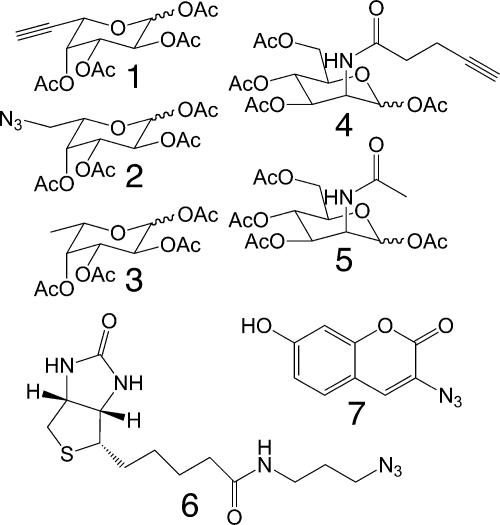
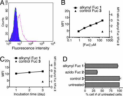
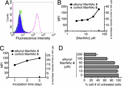
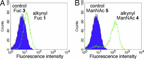
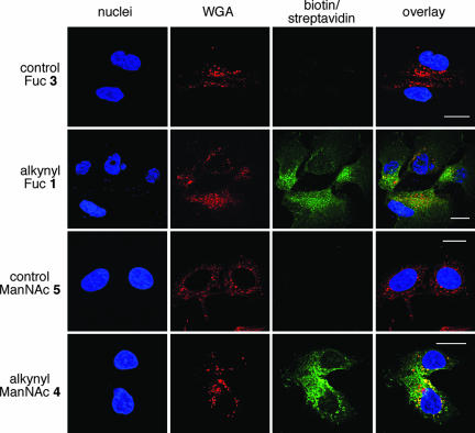
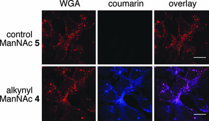
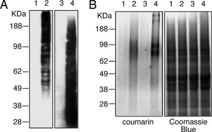
Developing tools for investigating the cellular activity of glycans will help to delineate the molecular basis for aberrant glycosylation in pathological processes such as cancer. Metabolic oligosaccharide engineering, which inserts sugar-reporting groups into cellular glycoconjugates, represents a powerful method for imaging the localization, trafficking, and dynamics of glycans and isolating them for glyco-proteomic analysis. Herein, we show that the alkyne-reporting group can be incorporated into cellular glycans. The alkyne group is a small, inert, bio-orthogonal handle that can be chemoselectively labeled by using the Cu(I) catalyzed [3 + 2] azide-alkyne cycloaddition, or click chemistry. Alkynyl sugar monomers, based on fucose (Fuc) and N-acetylmannosamine (ManNAc), were incorporated into fucosylated and sialylated glycans in several cancer cell lines, allowing for cell surface and intracellular visualization of glycoconjugates, as well as, observation of alkyne-bearing glycoproteins. Similarly to our previous results with an azido Fuc/alkynyl probe system, we demonstrated that click-activated fluorogenic probes are practical tools for efficiently and selectively labeling alkynyl-modified glycans. Because Fuc and sialic acid are terminal glycan residues with a notably increased presence in many tumors, we hope that our method will provide useful information about their roles in cancer and ultimately can be used for diagnostic and therapeutic purposes.


Conflict of interest statement

The authors declare no conflict of interest.

Figures

Scheme 1.
Scheme 1.
Modified sugar analogs and probes used in this study.
Fig. 1.
Fig. 1.
Analysis of cells labeled with Fuc analogs. (A) Flow cytometry analysis of Jurkat cells treated with Fuc analogs and labeled with biotin/fluorescein-conjugated streptavidin (filled histogram, untreated cells; green, cells treated with Fuc 3; pink, cells treated with alkynyl Fuc 1). (B) Dose-dependency of fucosyl glycan labeling by alkynyl Fuc 1 over 3 days. (C) Time course of fucosyl glycan labeling by 200 μM alkynyl Fuc 1. (D) Cell growth analysis after treatment with different Fuc analogs. Jurkat cells were grown in the presence of 200 μM each Fuc analog for 3 days before cell numbers were counted. The data represent the percentage of treated cells vs. untreated cells (n = 4).
Fig. 2.
Fig. 2.
Analysis of cell surface labeling of sialyl glycoconjugates. (A) Flow cytometry analysis of Jurkat cells labeled with alkynyl ManNAc (filled histogram, untreated cells; green, cells treated with control 5; pink, cells treated with alkynyl ManNAc 4). (B) Dose dependency of sialyl glycan labeling by alkynyl ManNAc 4 for 3 days. (C) Time course for labeling sialyl glycans by treatment with 25 μM alkynyl ManNAc 4. (D) Growth rate of Jurkat cells treated with different doses of alkynyl ManNAc 4 after 3 days.
Fig. 3.
Fig. 3.
Labeling of cell surface glycans by alkynyl sugar analogs and click-activated probe 3-azido-7-hydoxycoumarin (7). Shown is flow cytometry analysis of Jurkat cells labeled with 200 μM alkynyl Fuc 1 (A) or 25 μM alkynyl ManNAc 4 (B) for 3 days. The fluorescence intensity was detected after clicking on coumarin probe 7. Filled histogram, cells treated with sugar analog 3 or 5; open histogram, cell treated with alkynyl sugar 1 or 4.
Fig. 4.
Fig. 4.
Fluorescent imaging of fucosyl and sialyl glycoconjugates in cells. Confocal microscopy of Hep3B cells treated with 200 μM Fuc analogs or 25 μM ManNAc analogs. Cells were biotin-tagged and stained with streptavidin (fluorescein, green), WGA lectin (Alexa Fluor 594, red), and Hoechst 33342 (blue). (Scale bars: 20 μm.)
Fig. 5.
Fig. 5.
Visualization of sialyl glycoconjugates in cells using click-activated probe 7. Shown is confocal microscopy of coumarin-tagged Hep3B cells. Cells were treated with 25 μM ManNAc analogs 5 or 4 for 3 days, and then clicked with fluorogenic coumarin probe 7 (blue) and stained with WGA lectin (Alexa Fluor 594, red). (Scale bars: 20 μm.)
Fig. 6.
Fig. 6.
Detection of alkyne-labeled glycoproteins in cell extracts. Glycoproteins labeled with alkynyl sugars were derivatized and detected by immunoblotting of biotin tag (A) or fluorescent imaging of fluorogenic coumarin probe 7 (B). Protein extracts from cells grown with different sugars were analyzed (lane 1, control Fuc 3; lane 2, alkynyl Fuc 1; lane 3, control ManNAc 5; lane 4, alkynyl ManNAc 4). The protein gel (4–12%) was subsequently stained by Coomassie blue after fluorescent imaging, to verify equal protein loading.

Similar articles

Cited by

References

    1. Varki A, Cummings R, Esko JD, Freeze H, Hart GW, Marth J. Essentials of Glycobiology. Cold Spring Harbor, NY: Cold Spring Harbor Lab Press; 1999. pp. 1–635. - PubMed
    1. Axford JS. Biochim Biophys Acta. 1999;1455:219–229. - PubMed
    1. Dube DH, Bertozzi CR. Nat Rev Drug Discov. 2005;4:477–488. - PubMed
    1. Mackiewicz A, Mackiewicz K. Glycoconj J. 1995;12:241–247. - PubMed
    1. Meezan E, Wu HC, Black PH, Robbins PW. Biochemistry. 1969;8:2518–2524. - PubMed

Publication types