. 2007 Feb 20;104(8):2709-14.
doi: 10.1073/pnas.0608056104. Epub 2007 Feb 12.

Interaction between chromatin proteins MECP2 and ATRX is disrupted by mutations that cause inherited mental retardation

Affiliations

Interaction between chromatin proteins MECP2 and ATRX is disrupted by mutations that cause inherited mental retardation

Xinsheng Nan et al. Proc Natl Acad Sci U S A. .

Abstract

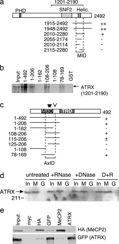
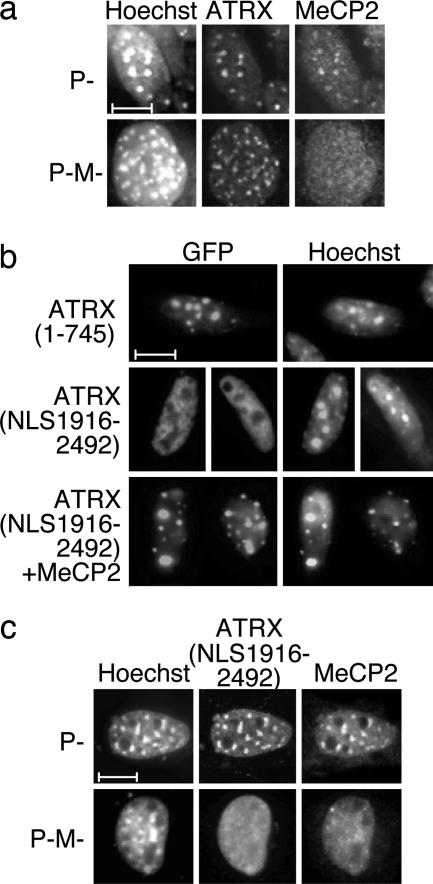
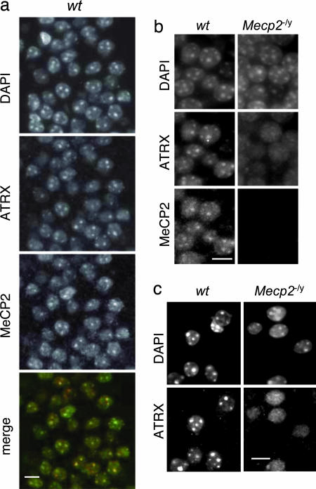
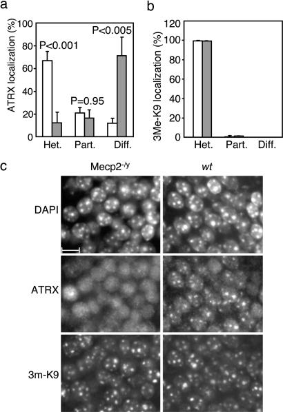
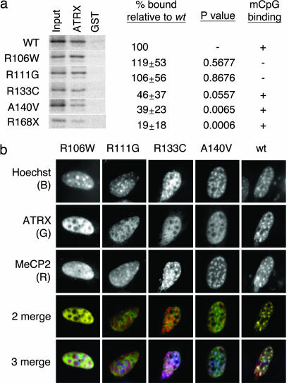
Mutations in the human methyl-CpG-binding protein gene MECP2 cause the neurological disorder Rett syndrome and some cases of X-linked mental retardation (XLMR). We report that MeCP2 interacts with ATRX, a SWI2/SNF2 DNA helicase/ATPase that is mutated in ATRX syndrome (alpha-thalassemia/mental retardation, X-linked). MeCP2 can recruit the helicase domain of ATRX to heterochromatic foci in living mouse cells in a DNA methylation-dependent manner. Also, ATRX localization is disrupted in neurons of Mecp2-null mice. Point mutations within the methylated DNA-binding domain of MeCP2 that cause Rett syndrome or X-linked mental retardation inhibit its interaction with ATRX in vitro and its localization in vivo without affecting methyl-CpG binding. We propose that disruption of the MeCP2-ATRX interaction leads to pathological changes that contribute to mental retardation.


Conflict of interest statement

The authors declare no conflict of interest.

Figures

Fig. 1.
Fig. 1.
MeCP2 interacts with ATRX. (a) Yeast two-hybrid assays identified a region of ATRX that interacts with MeCP2. The diagram depicts ATRX and its domains. Fragments 1915–2492 and 1948–2492 (amino acids numbered from the N terminus) were original clones from the library screen. Symbols ++, +, or − indicate the strength of the MeCP2 interaction based on expression of yeast HIS, ADE, and LacZ reporters (data not shown). The MeCP2 interaction domain (MID) is marked. (b) The ATRX-interaction region overlaps the MBD of MeCP2. The MeCP2 region that binds ATRX was identified by GST pulldown by using a deletion series of MeCP2-GST fusion proteins (for polyacrylamide gel of proteins, see SI Fig. 7) incubated with in vitro-translated [35S]-labeled ATRX fragment 1201–2190 (bracket; a). The input lane (Top) contained 25% of labeled protein used for the pull-down assay. (c) Summary of GST-pulldown showing presence (+) or absence (−) of an ATRX interaction leading to identification of an ATRX-interacting domain (AxID). Point mutations that inhibit ATRX binding (R133C, A140V, and R168X; see Fig. 5) are marked by arrows. (d) Native ATRX in nuclear extracts from brain is “pulled down” by immobilized full-length MeCP2. A MeCP2-GST fusion protein (M) or GST alone (G) were immobilized on glutathione beads and mixed with extract. Bound proteins and input (In; 10%) were separated and probed with anti-ATRX antibody on a Western blot. (e) CoIP of HA-tagged MeCP2 and GFP-fusion ATRX(NLS1201–2492) expressed transiently in mouse L cells. “Input” lane shows 10% of input amount. Antibodies used for coIP and blot visualization are labeled above and beside the figure, respectively.
Fig. 2.
Fig. 2.
ATRX localizes to heterochromatic foci in cultured mouse cells lacking DNA methylation, but exogenous MeCP2 can direct the ATRX C-terminal domain to heterochromatin in a DNA methylation-dependent manner. (a) Localization of endogenous ATRX in mouse fibroblasts is normally independent of MeCP2. Control P cells and the DNA methylation-deficient P-M cells were immunostained with anti-ATRX and anti-MeCP2 antibodies and counterstained with Hoechst 33258. (b) The C-terminal region of ATRX is targeted to heterochromatin by MeCP2. Constructs expressing GFP fused to the ATRX N terminus (1–745) or the ATRX C terminus (1916–2492), including an SV40 NLS, were transfected singly, or in combination with full-length MeCP2, into mouse L cells. (c) Targeting of the ATRX C terminus by MeCP2 depends on DNA methylation. ATRX (NLS1916–2492) and HA-MeCP2 were transiently coexpressed in control P or DNA methylation-deficient P-M cells. Localization was monitored by using anti-GFP and anti-HA antibodies. (Scale bars: 10 μm.)
Fig. 3.
Fig. 3.
Mislocalization of endogenous ATRX in the Mecp2-null mouse brain. (a) Immunostaining of ATRX, MeCP2, and DNA (DAPI) in wt hippocampus (CA1 region) confirms colocalization of MeCP2 and ATRX in neuronal nuclei (see yellow stain in merge). (b) Loss of ATRX localization in the Mecp2-null hippocampus. Speckled nuclear staining of ATRX coincides with MeCP2 foci in the wt dentate gyrus (for merged images, see SI Fig. 9), but dispersed nuclear ATRX staining is reproducibly seen in the Mecp2-null brain. (c) Immunostaining of ATRX in cortical neurons of wt and Mecp2−/y brains. (Scale bars: 10 μm.)
Fig. 4.
Fig. 4.
Heterochromatic foci persist in the Mecp2-null brain but do not sequester ATRX. (a) Dentate gyrus nuclei (≈100 per section, three brains per genotype) were graded for ATRX localization: predominantly heterochromatic staining (het.), heterochromatic plus diffuse staining (part.), and diffuse staining only (diff.). Significance of differences between wt (open bars) and mutant (shaded bars) are denoted by P values. (b) Dentate gyrus was also graded for staining with antibodies against trimethyl histone H3 lysine 9. (c) ATRX is delocalized in mutant nuclei that retain normal histone H3 lysine 9 trimethylation of heterochromatic foci. (Scale bar: 10 μm.)
Fig. 5.
Fig. 5.
Mutations in human in the MBD of human MeCP2 that cause mental retardation disturb the MeCP2–ATRX interaction without affecting methyl-CpG binding. (a) A GST fusion of ATRX (1915–2492) was immobilized and exposed to in vitro-translated [S35]-labeled MeCP2 (1–206) with the mutations indicated at the left. Shown is 10% of input, the amounts retained by ATRX (1915–2492) and by GST. Densitometric results (average of three experiments) are expressed relative to wt (% bound) together with P values (Student t test). The “mCpG binding” column summarizes the methyl-CpG binding as assayed in SI Fig. 15. (b) MeCP2 mutants A140V and R133C target heterochromatic foci but cannot direct ATRX to heterochromatin. HA-tagged full-length mutant MeCP2 forms were coexpressed with GFP-ATRX (NLS1916–2492) fusion proteins in mouse fibroblasts. Localization was monitored by immunostaining using anti-HA (MeCP2) and anti-GFP (ATRX) antibodies. In the merge, Hoechst, ATRX, and MeCP2 are blue, green, and red, respectively. R133C and A140V mutants colocalize with Hoechst bright spots (pink spots, triple merge), but ATRX staining is diffuse (absence of yellow spots, double merge). Note that MeCP2 R111G partially localizes to nucleoli.

Similar articles

Cited by

References

    1. Hong EJ, West AE, Greenberg ME. Curr Opin Neurobiol. 2005;15:21–28. - PubMed
    1. Jones PL, Veenstra GJ, Wade PA, Vermaak D, Kass SU, Landsberger N, Strouboulis J, Wolffe AP. Nat Genet. 1998;19:187–191. - PubMed
    1. Nan X, Campoy FJ, Bird A. Cell. 1997;88:471–481. - PubMed
    1. Nan X, Ng HH, Johnson CA, Laherty CD, Turner BM, Eisenman RN, Bird A. Nature. 1998;393:386–389. - PubMed
    1. Amir RE, Van den Veyver IB, Wan M, Tran CQ, Francke U, Zoghbi HY. Nat Genet. 1999;23:185–188. - PubMed

Publication types

MeSH terms