. 2007 Feb 20;104(8):2643-8.
doi: 10.1073/pnas.0606398104. Epub 2007 Feb 12.

A synthetic time-delay circuit in mammalian cells and mice

Affiliations

A synthetic time-delay circuit in mammalian cells and mice

Wilfried Weber et al. Proc Natl Acad Sci U S A. .

Abstract

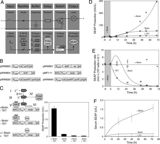
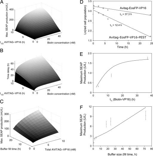
Time-delay circuitries in which a transcription factor processes independent input parameters can modulate NF-kappaB activation, manage quorum-sensing cross-talk, and control the circadian clock. We have constructed a synthetic mammalian gene network that processes four different input signals to control either immediate or time-delayed transcription of specific target genes. BirA-mediated ligation of biotin to a biotinylation signal-containing VP16 transactivation domain triggers heterodimerization of chimeric VP16 to a streptavidin-linked tetracycline repressor (TetR). At increasing biotin concentrations up to 20 nM, TetR-specific promoters are gradually activated (off to on, input signal 1), are maximally induced at concentrations between 20 nM and 10 microM, and are adjustably shut off at biotin levels exceeding 10 microM (on to off, input signal 2). These specific expression characteristics with a discrete biotin concentration window emulate a biotin-triggered bandpass filter. Removal of biotin from the culture environment (input signal 3) results in time-delayed transgene expression until the intracellular biotinylated VP16 pool is degraded. Because the TetR component of the chimeric transactivator retains its tetracycline responsiveness, addition of this antibiotic (input signal 4) overrides biotin control and immediately shuts off target gene expression. Biotin-responsive immediate, bandpass filter, and time-delay transcription characteristics were predicted by a computational model and have been validated in standard cultivation settings or biopharmaceutical manufacturing scenarios using trangenic CHO-K1 cell derivatives and have been confirmed in mice. Synthetic gene circuitries provide insight into structure-function correlations of native signaling networks and foster advances in gene therapy and biopharmaceutical manufacturing.


Conflict of interest statement

The authors declare no conflict of interest.

Figures

Fig. 1.
Fig. 1.
Design and functionality of the electronic and biologic time-delay circuits. (A) Electronic time-delay circuitry. Activating the switch triggers a current through the diode into the capacitor, which becomes charged and remains charged even when the switch is interrupted again. The capacitor's charge is slowly dissipated to the base of the resistor (b) where it triggers a current from collector (c) to emitter (e), resulting in activation of the output lamp. After complete capacitor draining the transistor switches back to off. In the synthetic biologic counterpart, the biotin input (I) results in VP16 biotinylation (II) and accumulation of biotinylated VP16 (III) in the cell buffer from which it is slowly degraded (IV). Biotinylated VP16 attaches to streptavidin fused with TetR, which binds specific tetO7 operator sites upstream of a minimal human cytomegalovirus promoter (Pmin), resulting in promoter activation (V) and production (VI) of the reporter SEAP. (B) Expression vectors used for construction of the biologic time-delay circuitry. at, AVITAG biotinylation signal; birA, E. coli biotin ligase; eosFP, photoswitchable fluorescent protein; pA, polyadenylation site; PCMV, human cytomegalovirus immediate early promoter; Pmin, minimal human cytomegalovirus promoter; PSV40, simian virus 40 promoter; pt, ubiquitinylation signal (PEST-sequence); sa, streptavidin; seap, human placental secreted alkaline phosphatase; tetO7, heptameric TetR-specific operator site; tetR, tetracycline-responsive repressor; vp16, activation domain of Herpes simplex viral protein 16. (C) Functional schematic of regulatory modules. In the presence of biotin (+Biotin; I) VP16 is biotinylated by BirA (II), accumulates in the cell (III), and activates (V) the minimal promoter (Pmin) by interaction with TetR-fused streptavidin (SA-TetR) bound to the tetO7 operator site in the absence of tetracycline, resulting in SEAP production (VI). Gene expression is reversed when biotinylated VP16 is degraded (IV). In the absence of biotin (−Biotin), VP16 is not biotinylated and the promoter remains silent. Upon addition of tetracycline (+TET) TetR-SA dissociates from tetO7 and the minimal promoter is not activated, irrespective of whether biotin is present or not. (D) In vitro and in silico validation of the time-delay circuitry. CHOTIME cells (50,000 cells per ml) were cultivated in standard biotin-containing (20 nM) medium for 9 h (gray shaded area) before switching (at t = 0 h) to biotin-free (−Biotin, circles), biotin-containing (+Biotin, 20 nM, crosses) or tetracycline-containing (+TET, 2 μg/ml, squares) medium. Time courses of SEAP production under these conditions include experimental data (symbols) and in silico predictions (lines). Details on the derivation of mathematical model, parameter values, and initial conditions are provided in supporting information (SI) Text and SI Tables 1 and 2. (E) Specific SEAP production rates calculated from finite differences of experimental data shown in D (symbols) and corresponding computational predictions (lines). (F) In vivo validation of the time-delay circuitry. Biotin-adapted (20 nM) CHOTIME cells were microencapsulated in alginate-poly-l-lysine-alginate microcapsules and i.p.-injected into mice followed by administration of biotin (100 μg/kg), tetracycline (100 mg/kg), or 0.9% NaCl as control (−Biotin). Serum samples were taken after 20, 40, and 72 h for quantification of SEAP production.
Fig. 2.
Fig. 2.
In silico and in vitro functional characterization of the biologic time-delay circuit and associated molecular building blocks. (A) Computational prediction of maximum SEAP production in a 72-h time interval after switching from biotin-containing medium (12-h cultivation) to biotin-free medium as a function of biotin concentration and AVITAG-VP16 half-life (t1/2 AVITAG-VP16). (B) Predicted time delays defined as the differences between the points in time of reaching 50% SEAP production rate after transcriptional shut-off caused by switching to tetracycline-containing and biotin-free medium, respectively. (C) Corresponding in silico-determined switching characteristics of the biologic time-delay circuitry as a function of the buffer size of total VP16 molecules and the buffer fill time (time of cultivation in 20 nM biotin medium). (D) Quantification of half-lives of different chimeric transactivators. CHO-K1 were transfected with plasmids pWW830 (PSV40-AVITAG-EosFP-VP16-pA) or pWW840 (PSV40-AVITAG-EosFP-VP16-PEST-pA) and cultivated for 48 h before switching the EosFP-fusion proteins from green to red by a 20-s UV (390 nm) light pulse. FACS analysis was used at the indicated time points to score the decrease in red fluorescence. (E) In vitro investigation of the impact of VP16 degradation kinetics on switching characteristics as represented by maximum SEAP production. CHO-K1 was transfected with pWW801, pWW804, pMF111, and either pWW800, pWW830, or pWW840 and cultivated for 2 h in the presence of biotin before switching to biotin-free conditions for 72 h and profiling of maximum SEAP production. Maximum SEAP production was correlated with the half-lives of AVITAG-VP16 variants (pWW800, 3.5 h; pWW840, 12.4 h; pWW830, 37.3 h) as predicted by the mathematical model (line). (F) In vitro investigation of the impact of the buffer size on switching characteristics represented by maximum SEAP production. CHO-K1 were transfected with plasmids pWW801, pWW804, pWW830, and pMF111 and cultivated in biotin-containing (20 nM) medium for the indicated periods before switching to biotin free conditions for another 48 h. Maximum SEAP production was determined and correlated with the time required to replenish the biotinylated VP16 buffer (line indicates model predictions).
Fig. 3.
Fig. 3.
Validation of biotin-dependent expression signaling for bioprocess applications and in vivo gene therapy studies. (A) Biotin dose–response study. CHO-K1 were transfected with plasmids pWW800, pWW801, pWW804, and pMF111 and cultivated in the presence of different biotin concentrations for 48 h before measuring SEAP production. The experimental data were used to estimate model parameters; simulation results are shown by the line. (B) Bioreactor compatibility of biotin-regulated gene expression. CHO-K1 cells, stably transfected with plasmids pWW800, pWW801, pWW804, and pMF111, were grown in a 1L BioWave reactor, and SEAP expression was induced at 48 h by addition of 100 nM biotin (gray area) and subsequently reversed at 125 h by the addition of 200 μM biotin. Lines indicate model predictions. (C) The same as B except that expression was reversed at 125 h by the addition of 2 μg/ml tetracycline. (D) Production of the toxic death domain of the human receptor-interacting protein (RipDD). CHO-K1 cells were transfected with plasmids pWW800, pWW801, pWW804, and pWW810 (PhCMV∗-1-RipDD-HIS6-pA) and cultivated in 20 nM biotin for the indicated time periods before Western blot-based RipDD quantification (IOD, integrated optical density) and assessment cell death (annexin and propidium iodide stainings) 48 h after transfection. (E) Validation of biotin-dependent signaling for in vivo gene expression control. CHO-K1 cells, stably transfected with plasmids pWW800, pWW801, pWW804, and pMF111, were microencapsulated and implanted into mice fed with a standard biotin-containing diet. Mice were injected with increasing biotin doses, and SEAP serum levels were assessed after 48 h.

Similar articles

Cited by

References

    1. Endy D. Nature. 2005;438:449–453. - PubMed
    1. Gibbs WW. Sci Am. 2004;290:74–81. - PubMed
    1. Hasty J, McMillen D, Collins JJ. Nature. 2002;420:224–230. - PubMed
    1. Sprinzak D, Elowitz MB. Nature. 2005;438:443–448. - PubMed
    1. Guido NJ, Wang X, Adalsteinsson D, McMillen D, Hasty J, Cantor CR, Elston TC, Collins JJ. Nature. 2006;439:856–860. - PubMed

Publication types

LinkOut - more resources