

Cognitive aging, executive function, and fractional anisotropy: a diffusion tensor MR imaging study
- PMID: 17296985
- PMCID: PMC7977408
Cognitive aging, executive function, and fractional anisotropy: a diffusion tensor MR imaging study
Abstract
Background and purpose: Fractional anisotropy (FA) is a useful measure of connectivity in the brain that can be derived from the diffusion tensor imaging (DTI) dataset. This study investigated the relationship between FA and selected measures of cognition across a broad age group to explore a possible structural basis for cognitive changes with age.
Methods: FA images were generated from DTI data acquired at 1.5T in 87 healthy subjects (age range, 20-73 years). Relationships between a range of cognitive measures and FA were explored using regional and voxel-based analysis.
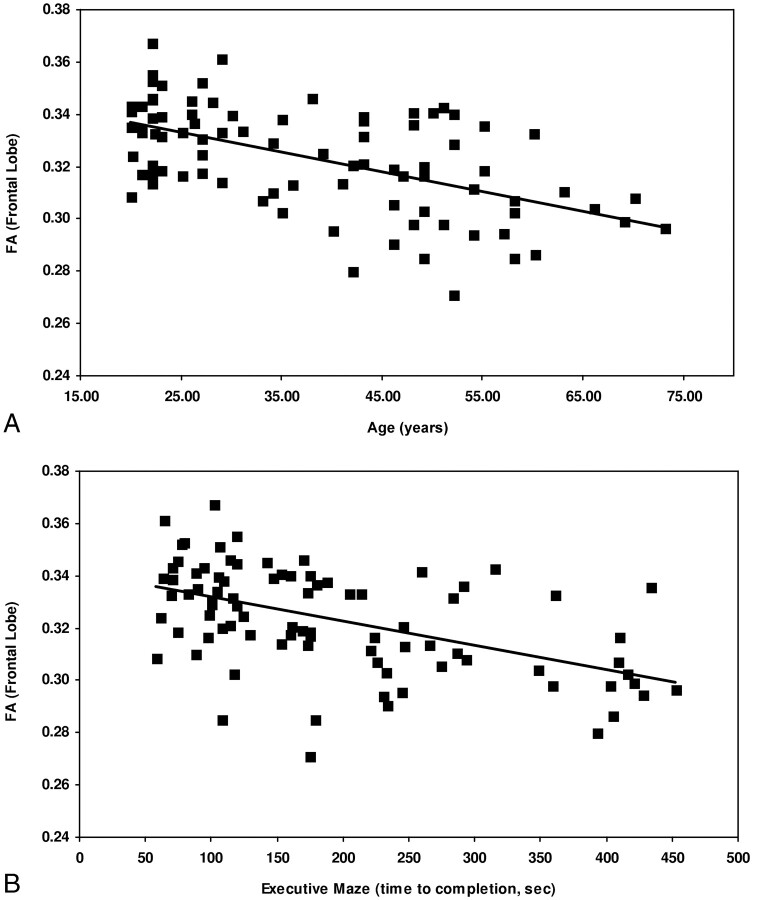
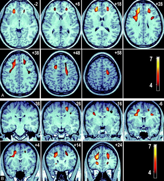
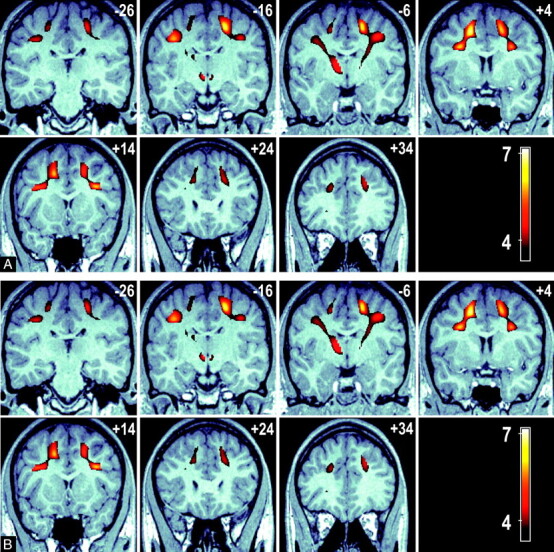
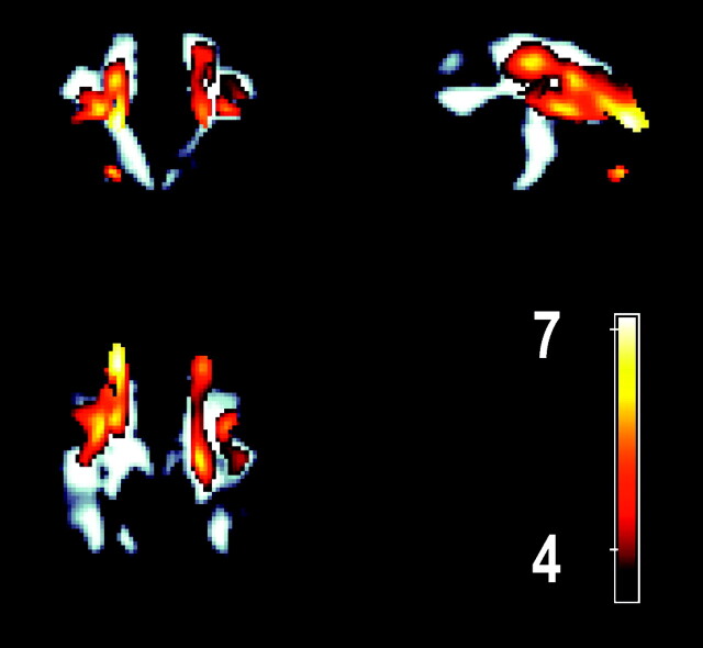
Results: Age and regional average FA were significantly associated in the frontal, parietal, and temporal lobes but not in the occipital lobe. This negative relationship was especially prominent in the prefrontal regions of the frontal lobe, where FA declined at a rate of approximately 3% per decade. Decreased FA in the frontal, temporal, and parietal lobes was associated with poorer cognitive performance in executive maze and in an attention-switching task. A voxel-level analysis of these data revealed that the executive function-FA association was particularly strong and regionally delineated over 2 continuous, bilateral areas extending from the prefrontal cortex to the parietal lobe, with projections to the anterior portions of the thalamus.
Conclusions: We demonstrate a relationship between FA and a measure of executive function-a core cognitive component that is a key feature of cognitive aging. We propose that that FA may provide an early means for the detection of age-related cognitive change and suggest a need for prospective data to explore this association.
Figures
References
-
- Basser PJ, Mattiello J, LeBihan D. Estimation of the effective self-diffusion tensor from the NMR spin echo. J Magn Reson B 1994;103:247–54 - PubMed
-
- Hahn EL. Spin echoes. Phys Rev 1950;80:580
-
- Ciccarelli O, Werring DJ, Barker GJ, et al. A study of the mechanisms of normal-appearing white matter damage in multiple sclerosis using diffusion tensor imaging–evidence of Wallerian degeneration. J Neurol 2003;250:287–92 - PubMed
-
- Fellgiebel A, Wille P, Muller MJ, et al. Ultrastructural hippocampal and white matter alterations in mild cognitive impairment: a diffusion tensor imaging study. Dement Geriatr Cogn Disord 2004;18:101–08 - PubMed
MeSH terms
LinkOut - more resources
Full Text Sources
Medical