

Hyperintensity of the precentral gyral subcortical white matter and hypointensity of the precentral gyrus on fluid-attenuated inversion recovery: variation with age and implications for the diagnosis of amyotrophic lateral sclerosis
- PMID: 17296988
- PMCID: PMC7977425
Hyperintensity of the precentral gyral subcortical white matter and hypointensity of the precentral gyrus on fluid-attenuated inversion recovery: variation with age and implications for the diagnosis of amyotrophic lateral sclerosis
Abstract
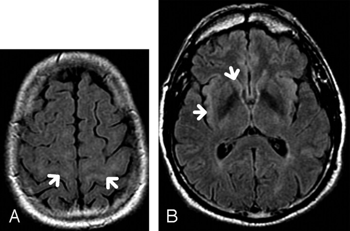
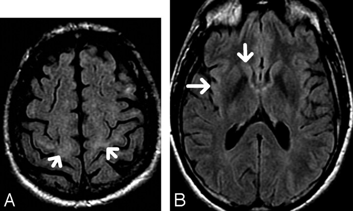
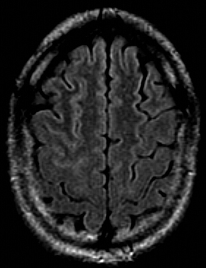
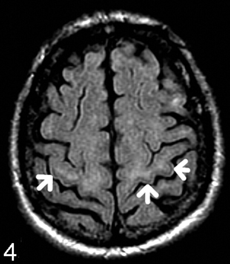
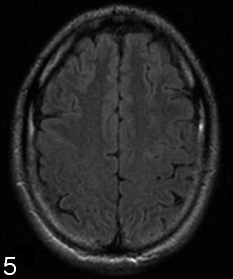
Background and purpose: Hyperintensity of the subcortical white matter (SWM) of the precentral gyrus and hypointensity of the precentral gyrus gray matter (PGGM) on fluid-attenuated inversion recovery (FLAIR) are described as potentially useful diagnostic findings in amyotrophic lateral sclerosis (ALS). A detailed study of the prevalence of these findings in various age groups has not been described.
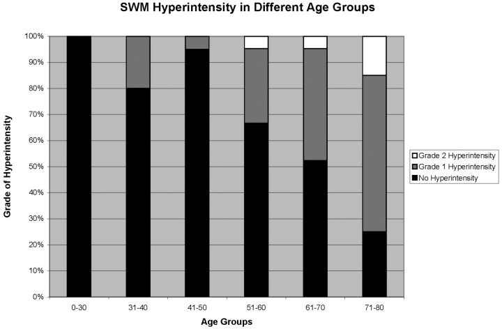
Methods: One hundred twenty-two patients underwent axial FLAIR brain examinations as part of either hearing loss or tinnitus evaluation. Examinations were randomly selected to reflect an even spread through the decades from ages 15 to 78 years and were reviewed by 2 readers, blinded to patient's age and sex, for the presence/absence of the above 2 signs. If SWM hyperintensity was present, it was graded as intense as caudate nucleus (grade 1) or insula (grade 2).
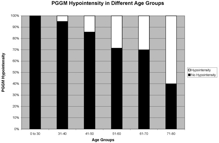
Results: We identified 32 cases of grade 1 and 5 cases of grade 2 SWM hyperintensity, and 28 cases of PGGM hypointensity. Both signs showed significant Spearman correlation with increasing age (r = 0.55, P < .001 for grade 1, r = 0.45, P < .001 for grade 2 SWM hyperintensity, r = 0.45, P < .001 for PGGM hypointensity). Analysis of variance showed there was a significant difference between the different age groups (P < .001) for both signs. Grading of the SWM and PGGM signals were highly reproducible with very good interobserver agreement (r = 0.88, P < .001, and r = 0.97, P < .001, respectively).
Conclusion: This study suggests a statistically significant relationship between increasing age and the frequency of precentral gyrus SWM hyperintensity and PGGM hypointensity on FLAIR, and reinforces previous reports that these signs can be seen in patients who do not have ALS.
Figures
Similar articles
-
Hyperintensity of the corticospinal tract on FLAIR: A simple and sensitive objective upper motor neuron degeneration marker in clinically verified amyotrophic lateral sclerosis.J Neurol Sci. 2016 Aug 15;367:177-83. doi: 10.1016/j.jns.2016.06.005. Epub 2016 Jun 3. J Neurol Sci. 2016. PMID: 27423585
-
The diagnostic utility of FLAIR imaging in clinically verified amyotrophic lateral sclerosis.J Magn Reson Imaging. 2003 May;17(5):521-7. doi: 10.1002/jmri.10293. J Magn Reson Imaging. 2003. PMID: 12720261
-
Hyperintensity of the middle cerebellar peduncles on fluid-attenuated inversion recovery imaging: variation with age and implications for the diagnosis of multiple system atrophy.AJNR Am J Neuroradiol. 2006 Nov-Dec;27(10):2146-8. AJNR Am J Neuroradiol. 2006. PMID: 17110685 Free PMC article.
-
[Objective markers for upper motor neuron involvement in amyotrophic lateral sclerosis].Brain Nerve. 2007 Oct;59(10):1053-64. Brain Nerve. 2007. PMID: 17969345 Review. Japanese.
-
[MR imaging of the brain of amyotrophic lateral sclerosis].Rinsho Shinkeigaku. 1995 Dec;35(12):1554-6. Rinsho Shinkeigaku. 1995. PMID: 8752461 Review. Japanese.
Cited by
-
Mapping cortical degeneration in ALS with magnetization transfer ratio and voxel-based morphometry.PLoS One. 2013 Jul 9;8(7):e68279. doi: 10.1371/journal.pone.0068279. Print 2013. PLoS One. 2013. PMID: 23874570 Free PMC article.
-
Five-class differential diagnostics of neurodegenerative diseases using random undersampling boosting.Neuroimage Clin. 2017 Jun 12;15:613-624. doi: 10.1016/j.nicl.2017.06.012. eCollection 2017. Neuroimage Clin. 2017. PMID: 28664032 Free PMC article.
-
Value of fluid-attenuated inversion recovery MRI data analyzed by the lesion segmentation toolbox in amyotrophic lateral sclerosis.J Magn Reson Imaging. 2019 Aug;50(2):552-559. doi: 10.1002/jmri.26577. Epub 2018 Dec 19. J Magn Reson Imaging. 2019. PMID: 30569457 Free PMC article.
-
Impaired structural motor connectome in amyotrophic lateral sclerosis.PLoS One. 2011;6(9):e24239. doi: 10.1371/journal.pone.0024239. Epub 2011 Sep 2. PLoS One. 2011. PMID: 21912680 Free PMC article.
-
Magnetization transfer imaging demonstrates a distributed pattern of microstructural changes of the cerebral cortex in amyotrophic lateral sclerosis.AJNR Am J Neuroradiol. 2011 Apr;32(4):704-8. doi: 10.3174/ajnr.A2356. Epub 2011 Mar 24. AJNR Am J Neuroradiol. 2011. PMID: 21436337 Free PMC article. Clinical Trial.
References
-
- Zhang L, Ulug AM, Zimmerman RD, et al. The diagnostic utility of FLAIR imaging in clinically verified amyotrophic lateral sclerosis. J Magn Reson Imaging 2003 May;17:521–7 - PubMed
-
- Hecht MJ, Fellner F, Fellner C, et al. MRI-FLAIR images of the head show corticospinal tract alterations in ALS patients more frequently than T2-, T1- and proton-density weighted images. J Neurol Sci 2001;186:37–44 - PubMed
-
- Chan S, Kaufmann P, Shungu DC, et al. Amyotrophic lateral sclerosis and primary lateral sclerosis: evidence-based diagnostic evaluation of the upper motor neuron. Neuroimaging Clin N Am 2003 May;13:307–26 - PubMed
-
- Hecht MJ, Fellner F, Fellner C, et al. Hyperintense and hypointense MRI signals of the precentral gyrus and corticospinal tract in ALS: a follow-up examination including FLAIR images. J Neurol Sci 2002;199:59–65 - PubMed
MeSH terms
LinkOut - more resources
Full Text Sources
Medical
Miscellaneous