

Whole-brain and regional brain atrophy in amyotrophic lateral sclerosis
- PMID: 17296989
- PMCID: PMC7977419
Whole-brain and regional brain atrophy in amyotrophic lateral sclerosis
Abstract
Background and purpose: Recent evidence from neuropsychologic and neuroimaging studies suggests that central nervous system involvement in amyotrophic lateral sclerosis (ALS) extends beyond motor neurons. Our purpose was to obtain measures of global and regional atrophy in nondemented patients with ALS to assess subtle structural brain changes.
Methods: MR images, acquired from 16 patients and 9 healthy subjects (HS), were processed by using the Structural Imaging Evaluation of Normalized Atrophy (SIENA) software to estimate whole-brain atrophy measures and the voxel-based morphometry (VBM) method to highlight the selective volumetric decrease of single cerebral areas. In addition, each subject underwent a neuropsychologic examination.
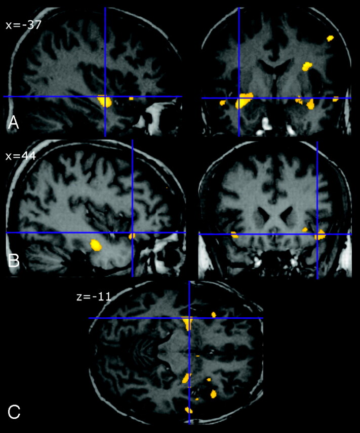
Results: In patients with ALS, brain parenchymal fraction was slightly lower compared with HS (P = .012), and seemed to be related to the presence of cognitive impairment. Patients showed a gray matter volume decrease in several frontal and temporal areas bilaterally (P < .001 uncorrected) compared with HS, with a slight prevalence in the right hemisphere. No volume reduction in primary motor cortices of patients was detected. Performances on Symbol Digit Modalities Test were significantly worse in patients compared with HS (P = .025).
Conclusions: The presence of mild whole-brain volume loss and regional frontotemporal atrophy in patients with ALS could explain the presence of cognitive impairment and confirms the idea of ALS as a degenerative brain disease not confined to motor system.
Figures
Comment in
-
Regional assessment of brain atrophy: a novel approach to achieve a more complete picture of tissue damage associated with central nervous system disorders?AJNR Am J Neuroradiol. 2007 Feb;28(2):260-1. AJNR Am J Neuroradiol. 2007. PMID: 17296990 Free PMC article. Review. No abstract available.
-
Computer-based 3D MR imaging analysis in amyotrophic lateral sclerosis: common and specific factors among studies.AJNR Am J Neuroradiol. 2007 Oct;28(9):1626; author reply 1627. doi: 10.3174/ajnr.A0681. Epub 2007 Sep 20. AJNR Am J Neuroradiol. 2007. PMID: 17885226 Free PMC article. No abstract available.
Similar articles
-
Gray matter and white matter changes in non-demented amyotrophic lateral sclerosis patients with or without cognitive impairment: A combined voxel-based morphometry and tract-based spatial statistics whole-brain analysis.Brain Imaging Behav. 2018 Apr;12(2):547-563. doi: 10.1007/s11682-017-9722-y. Brain Imaging Behav. 2018. PMID: 28425061
-
Structural and functional evaluation of cortical motor areas in Amyotrophic Lateral Sclerosis.Exp Neurol. 2012 Mar;234(1):169-80. doi: 10.1016/j.expneurol.2011.12.024. Epub 2011 Dec 27. Exp Neurol. 2012. PMID: 22226599
-
Relationship between Clinical Parameters and Brain Structure in Sporadic Amyotrophic Lateral Sclerosis Patients According to Onset Type: A Voxel-Based Morphometric Study.PLoS One. 2017 Jan 17;12(1):e0168424. doi: 10.1371/journal.pone.0168424. eCollection 2017. PLoS One. 2017. PMID: 28095425 Free PMC article.
-
Structural explanation of poor prognosis of amyotrophic lateral sclerosis in the non-demented state.Eur J Neurol. 2017 Jan;24(1):122-129. doi: 10.1111/ene.13163. Epub 2016 Oct 18. Eur J Neurol. 2017. PMID: 27753163 Free PMC article.
-
The anatomy of cognitive impairment in amyotrophic lateral sclerosis: more than frontal lobe dysfunction.Cortex. 2012 Feb;48(2):166-82. doi: 10.1016/j.cortex.2011.02.004. Epub 2011 Feb 12. Cortex. 2012. PMID: 21396632 Review.
Cited by
-
The cortical signature of amyotrophic lateral sclerosis.PLoS One. 2012;7(8):e42816. doi: 10.1371/journal.pone.0042816. Epub 2012 Aug 6. PLoS One. 2012. PMID: 22880116 Free PMC article.
-
Brain atrophy and neuropsychological outcome after treatment of ruptured anterior cerebral artery aneurysms: a voxel-based morphometric study.Neuroradiology. 2009 Nov;51(11):711-22. doi: 10.1007/s00234-009-0552-5. Epub 2009 Jul 1. Neuroradiology. 2009. PMID: 19568738 Clinical Trial.
-
Modeling seeding and neuroanatomic spread of pathology in amyotrophic lateral sclerosis.Neuroimage. 2022 May 1;251:118968. doi: 10.1016/j.neuroimage.2022.118968. Epub 2022 Feb 7. Neuroimage. 2022. PMID: 35143975 Free PMC article.
-
Amyotrophic Lateral Sclerosis: A Neurodegenerative Motor Neuron Disease With Ocular Involvement.Front Neurosci. 2020 Sep 25;14:566858. doi: 10.3389/fnins.2020.566858. eCollection 2020. Front Neurosci. 2020. PMID: 33071739 Free PMC article. Review.
-
Microstructural changes across different clinical milestones of disease in amyotrophic lateral sclerosis.PLoS One. 2015 Mar 20;10(3):e0119045. doi: 10.1371/journal.pone.0119045. eCollection 2015. PLoS One. 2015. PMID: 25793718 Free PMC article.
References
-
- Rowland LP, Shneider NA. Amyotrophic lateral sclerosis. N Engl J Med 2001;344:1688–700 - PubMed
-
- Lomen-Hoerth C, Murphy J, Langmore S, et al. Are amyotrophic lateral sclerosis patients cognitively normal? Neurology 2003;60:1094–97 - PubMed
-
- Ringholz GM, Appel SH, Bradshaw M, et al. Prevalence and patterns of cognitive impairment in sporadic ALS. Neurology 2005;65:586–90 - PubMed
-
- Strong MJ, Lomen-Hoerth C, Caselli RJ, et al. Cognitive impairment, frontotemporal dementia, and the motor neuron diseases. Ann Neurol 2003;54:S20–23 - PubMed
MeSH terms
LinkOut - more resources
Full Text Sources
Medical
Miscellaneous