

Imaging degeneration of the substantia nigra in Parkinson disease with inversion-recovery MR imaging
- PMID: 17297002
- PMCID: PMC7977418
Imaging degeneration of the substantia nigra in Parkinson disease with inversion-recovery MR imaging
Abstract
Background and purpose: Visualizing with MR imaging and obtaining quantitative indexes of degeneration of the substantia nigra in Parkinson disease have been long-sought goals. We investigated the potential role of area and T1 contrast measurements in differentiating patients from controls and their age-related changes.
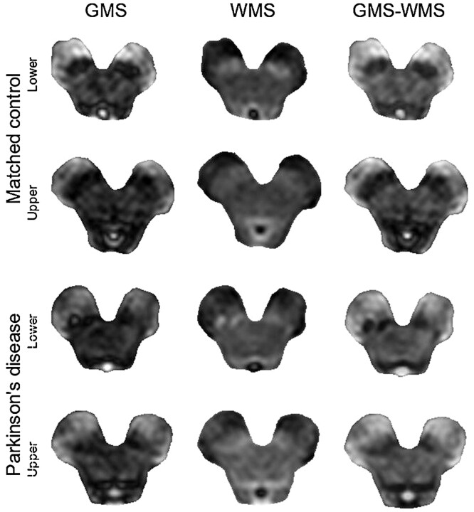
Methods: Eight patients with Parkinson disease, 8 age-matched controls, and 8 young controls were imaged. We obtained the pixel-wise difference between 2 sets of inversion-recovery images, acquired parallel to the bicommissural plane, with different inversion times. Pixel-intensity ratios between lateral and medial nigral regions, and nigral area and substantia-nigra/midbrain area ratios were computed.
Results: Compared with that of controls, loss of substantia nigra was evident in patients, its borders taking a smoother and more irregular appearance. Patients were characterized by a lateral-to-medial gradient, due to reduced hypointensity of the lateral portion of the substantia nigra and relative sparing of its medial portion. The visible nigral area was significantly smaller in patients compared with matched controls (P = .04). The substantia nigra/midbrain area ratio enabled considerably better separation (P = .0001). The lateral/medial pixel-intensity ratio was significantly higher in patients compared with matched controls (P = .01) and in young controls compared with age-matched controls (P = .01).
Conclusion: Inversion-recovery sequences may provide a convenient way to visualize nigral degeneration. Relative area and pixel-intensity measurements may integrate other techniques (such as diffusion-tensor imaging on nigrostriatal pathways) in the neuroradiologic diagnosis and follow-up of Parkinson disease by quantitatively assessing the degeneration of the substantia nigra.
Figures


References
-
- Elkeslassy A, Miaux Y, Martin-Duverneuil N, et al. MRI of degenerative extrapyramidal syndromes: Parkinson disease, progressive supranuclear palsy and multiple system atrophy. J Neuroradiol. 1996;23:157–63 - PubMed
-
- Ravina B, Eidelberg D, Ahlskog JE, et al. The role of radiotracer imaging in Parkinson disease. Neurology 2005;64:208–15 - PubMed
-
- Ordidge RJ, Gorell JM, Deniau JC, et al. Assessment of relative brain iron concentrations using T2-weighted and T2*-weighted MRI at 3 Tesla. Magn Reson Med 1994;32:335–41 - PubMed
-
- Gorell JM, Ordidge RJ, Brown GG, et al. Increased iron-related MRI contrast in the substantia nigra in Parkinson’s disease. Neurology 1995;45:1138–43 - PubMed
-
- Graham JM, Paley MN, Grunewald RA, et al. Brain iron deposition in Parkinson’s disease imaged using the PRIME magnetic resonance sequence. Brain 2000;123:2423–31 - PubMed
Publication types
MeSH terms
LinkOut - more resources
Full Text Sources
Other Literature Sources
Medical