

Case Reports
Imaging cerebral amyloid angiopathy with susceptibility-weighted imaging
Affiliations
- PMID: 17297004
- PMCID: PMC7977403
Item in Clipboard
Case Reports
Imaging cerebral amyloid angiopathy with susceptibility-weighted imaging
AJNR Am J Neuroradiol.
2007 Feb.
Abstract
Gradient-echo (GE) imaging is recognized as a means to detect hemorrhagic changes in cerebral amyloid angiopathy (CAA). However, almost 25% of patients with CAA do not show microhemorrhages on T2* GE imaging. We applied a new imaging method, susceptibility weighted imaging (SWI), to evaluate the presence of microhemorrhages. In a suspected case of CAA, where cognitive effects are also present, we show that SWI is much more sensitive in detecting microhemorrhages than conventional methods.
Figures
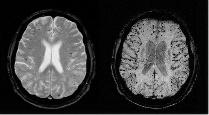
Axial gradient-echo T2*-weighted image (left; 800/26 ms [TR/TE], 5-mm section thickness, Nx = 256, Ny = 154) shows some low-signal-intensity foci associated with CAA. Corresponding SWI image (right; 85/35 ms [TR/TE], α = 20°, Nx = 512, Ny = 256, with a resolution of 0.5 mm × 0.5 mm × 2.0 mm, projected over 8 mm) shows many more associated low-signal-intensity foci.
References
-
- Reichenbach JR, Venkatesan R, Schillinger DJ, et al. Small vessels in the human brain: MR venography with deoxyhemoglobin as an intrinsic contrast agent. Radiology 204 ;1997. :272–77 - PubMed
-
- Reichenbach JR, Jonetz-Mentzel L, Fitzek C, et al. High-resolution blood oxygen-level dependent MR venography (HRBV): a new technique. Neuroradiology 43 ;2001. :364–69 - PubMed
-
- Sehgal V, DelProposto Z, Haacke EM et al. Clinical applications of neuroimaging with susceptibility-weighted imaging. J Magn Reson Imaging 22 ;2005. :439–50 - PubMed
-
- Tong KA, Ashwal S, Holshouser S, et al. Improved detection of hemorrhagic shearing lesions in children with post-traumatic diffuse axonal injury: initial results. Radiology 227 ;2003. :332–39 - PubMed
-
- Greenberg SM, O’Donnell HC, Schaefer PW, et al. MR imaging detection of new hemorrhages: potential marker of progression in cerebral amyloid angiopathy. Neurology 53 ;1999. :1135–38 - PubMed
Publication types
MeSH terms
Grants and funding
LinkOut - more resources
Full Text Sources
Other Literature Sources
Medical