

Case Reports
Intracranial dural fistulas with exclusive perimedullary drainage: the need for complete cerebral angiography for diagnosis and treatment planning
Affiliations
- PMID: 17297010
- PMCID: PMC7977392
Item in Clipboard
Case Reports
Intracranial dural fistulas with exclusive perimedullary drainage: the need for complete cerebral angiography for diagnosis and treatment planning
AJNR Am J Neuroradiol.
2007 Feb.
Abstract
Three patients are presented with slowly progressive tetraparesis caused by an intracranial dural arteriovenous fistula with exclusive perimedullary venous drainage. MR imaging showed a swollen cervicothoracic cord with central myelopathy and dilated perimedullary veins. Bilateral vertebral angiography initially failed to demonstrate the fistulas, and diagnosis was established with external carotid angiography. All 3 patients were successfully treated with glue embolization, 1 after failed surgical exploration. Angiographic cure of the fistula resulted in clinical cure in 1 patient and stabilization in 2 patients.
Figures
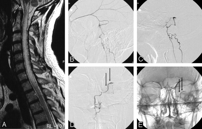
A 58-year-old man with progressive cervical cord dysfunction. A, MR image shows a swollen cervical cord with edema and central myelopathy and dilated perimedullary veins. B, Lateral view of a selective angiogram of the left middle meningeal artery demonstrates a dural fistula, with drainage to the perimedullary veins. C and D, Lateral (C) and anteroposterior (D) angiograms of the squamous branch of the middle meningeal artery contributing to the fistula show the beginning of the draining vein (arrows). Glue was injected from this position. E, Anteroposterior radiograph after glue injection shows glue in the draining vein (arrows).
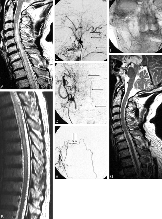
A 65-year-old man with progressive cord dysfunction. A and B, MR image shows central myelopathy in the cervicothoracic cord, with engorged perimedullary veins. C and D, Lateral (C) and anteroposterior (D) right external carotid angiograms demonstrate a fistula with drainage to the perimedullary veins (arrows). E, Anteroposterior projection of selective injection of a branch of the stylomastoid artery supplying the fistula. The proximal part of draining vein is marked with arrows. F, Radiograph, same as in E, shows glue in the proximal draining vein (arrows). G, Normal findings on MR image 1 year later.
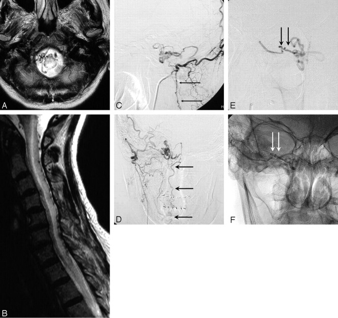
A 72-year-old woman with progressive tetraparesis. A and B, MR images show a convolute of veins at the level of the foramen magnum, central myelopathy and edema from the medulla oblongata to T6, and dilated posterior medullary veins. C and D, Lateral (C) and anteroposterior (D) right occipital artery angiograms show a dural fistula at the skull base, draining via a convolute of paramedullary veins into the posterior medullary veins (arrows). E, Injection via microcatheter shows the proximal draining vein (arrows). F, Radiograph, same as E, shows glue cast in the proximal draining vein (arrows).
References
-
- Mullan S. Reflections upon the nature and management of intracranial and intraspinal vascular malformations and fistulae. J Neurosurg 1994;80:606–16 - PubMed
-
- Thompson BG, Doppman JL, Oldfield EH. Treatment of cranial dural arteriovenous fistulae by interruption of leptomeningeal venous drainage. J Neurosurg 1994;80:617–23 - PubMed
-
- Jellema K, Sluzewski M, van Rooij WJ, et al. Embolization of spinal dural arteriovenous fistulas: importance of occlusion of the draining vein. J Neurosurg Spine 2005;2:580–83 - PubMed
-
- Gobin YP, Rogopoulos A, Aymard A, et al. Endovascular treatment of intracranial dural arteriovenous fistulas with spinal perimedullary venous drainage. J Neurosurg 1992;77:718–23 - PubMed
Publication types
MeSH terms
LinkOut - more resources
Full Text Sources