. 2007 Jul 19;26(33):4825-32.
doi: 10.1038/sj.onc.1210282. Epub 2007 Feb 12.

HEI10 negatively regulates cell invasion by inhibiting cyclin B/Cdk1 and other promotility proteins

Affiliations

HEI10 negatively regulates cell invasion by inhibiting cyclin B/Cdk1 and other promotility proteins

M K Singh et al. Oncogene. .

Abstract

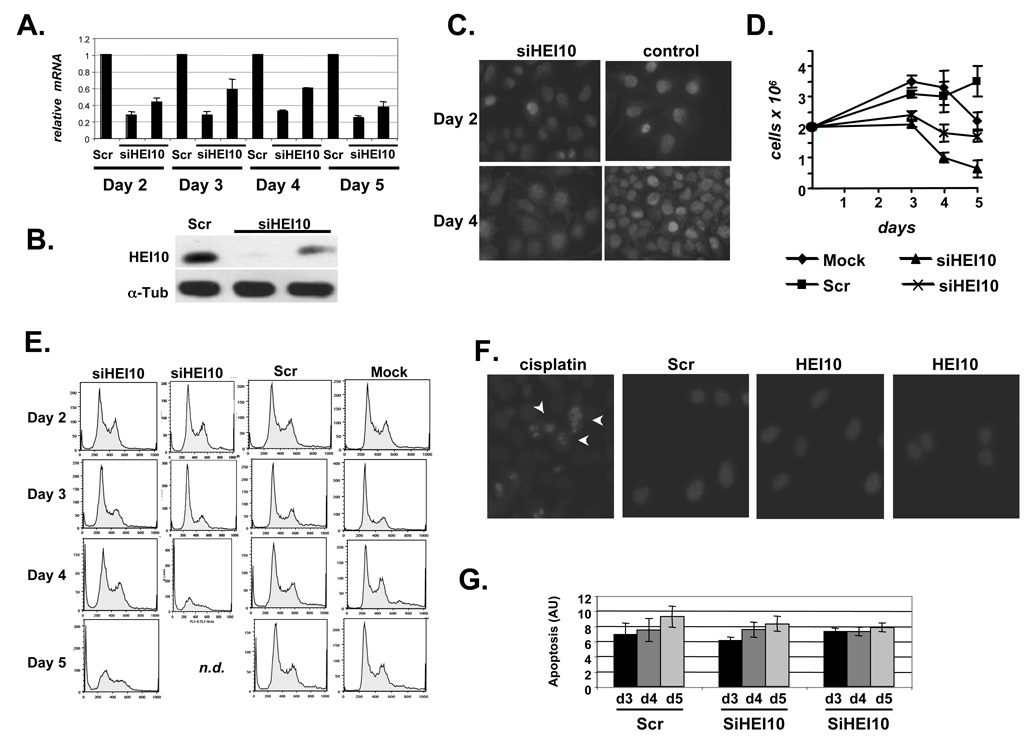
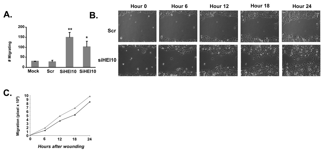
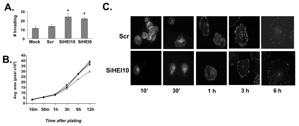
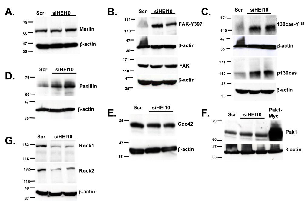
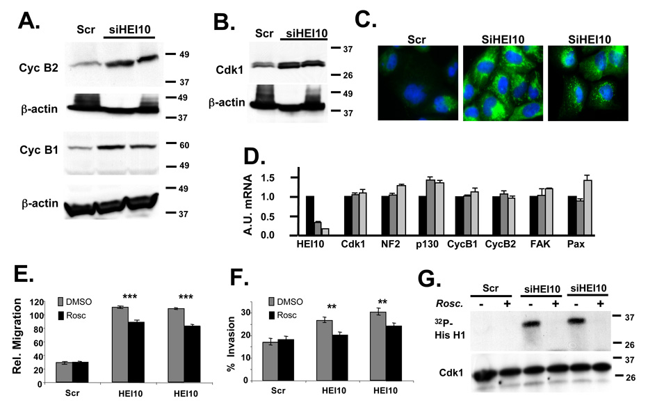
Human enhancer of invasion, clone 10 (HEI10) (CCNB1IP1) was first described as a RING-finger family ubiquitin ligase that regulates cell cycle by interacting with cyclin B and promoting its degradation. Subsequently, other studies suggested specific upregulation of HEI10 in metastatic melanoma and demonstrated direct interaction between HEI10 and the tumor suppressor Merlin, encoded by the neurofibromatosis 2 gene. These and other results led us to hypothesize that HEI10 also influences the processes of cell migration and metastasis. We here show that cells with depleted HEI10 both migrate more rapidly and invade more effectively than control cells. HEI10 depletion post-transcriptionally increases the expression of a group of promotility regulatory proteins including p130Cas, paxillin, Cdk1 and cyclin B2, but excluding Merlin. Among these, only inhibition of Cdk1/cyclin B activity specifically reversed the motility and invasion of HEI10-depleted cells. Finally, HEI10 is abundantly transcribed in many human tissues, and particularly abundant in some tumor cell lines, suggesting that it may be commonly involved in coordinating cell cycle with cell migration and invasion.


Figures

Figure 1
Figure 1. HEI10 depletion inhibits cell proliferation
A. HEI10 depetion by siRNA was analyzed by RT q-PCR in all experiments described in this study. Results shown are normalized to levels of RNApolR2F. B. Western blot showing HEI10 depleted by two siRNAs, in reference to a-tubulin loading control. C. Immunofluorescence with antibody to HEI10 cells treated with siRNAs to HEI10 and control. Results were consistent between two different siRNAs to HEI10 (second not shown). Note increased number of cells in control panel at day 4 (compare to C). D. Proliferation of cell lines mock-transfected, or transfected with control siRNAs (Scr), or two siRNAs targeting HEI10. E. FACS analysis of cells treated with two HEI10-targeted siRNAs, control siRNAs, or mock-transfected, at 2, 3, 4, and 5 days after treatment. n.d., not determined because of reduced cell numbers. F. Hoechst 33342 staining of nuclei of cells treated with Scr or HEI0-directed siRNAS for 4 days, or with cisplatin (arrows indicate pycnotic nuclei). G. Relative apoptotic index produced by APOPercentage assay at days 3, 4, and 5 after HEI10 depletion. Differences between HEI10- and Scr-depleted cells are not statistically significant.
Figure 2
Figure 2. HEI10 depletion increases cell migration
A. Migration of U2OS cells treated with two different HEI10-directed (siHEI10) or control (Scr) siRNAs through wells of a Boyden chamber. This experiment was performed 3 times in triplicate; numbers shown reflect average values. Similar results were obtained with siRNA treated MCF7 cells (not shown). Statistical analysis of Scr versus the two HEI10-targeted siRNAs indicates **P < 0.001 and *P< 0.01 respectively. B. Comparison of “wound” closure over 24 hours following scratching of a monolayer of cells treated 48 hours previously with two different HEI10-directed (siHEI10) or control (Scr) siRNAs. C. Quantitation of results in B. HEI10-depleted cells (squares, gray line) migrate more rapidly than Scr-depleted cells (diamonds, black line), P < 0.005 or better at all time points.
Figure 3
Figure 3. HEI10 depletion increases cell invasion and reattachment
A Migration of U2OS cells treated with two different HEI10-directed (siHEI10) or control (Scr) siRNAs through Boyden chamber wells coated with Matrigel, versus uncoated wells. Experiment was performed 3 times in triplicate; numbers shown reflect average values. Statistical analysis assigns difference between Scr and HEI10-directed siRNAs as *P < 0.01 in each case. B. U2OS cells treated with two HEI10-directed (siHEI10) or control (Scr) siRNAs for 48 hours were trypsinized, then replated on tissue culture plastic, and fixed at time points up to 24 hours. Outlines of cells (stained with antibody to paxillin) were traced using the Metamorph software program, with 100 cells counted for each time point in each of 3 experiments. Statistical confidence for difference HEI10- versus Scr-depleted is P <0.02 at 6 hrs, and P <0.01 at 12 hours. Black lines with circle or diamond, HEI10 depletion; gray line with triangle, Scr depletion. C. Representative cells analyzed for the data graphed in 3B.
Figure 4
Figure 4. HEI10 negatively regulates p130Cas, paxillin, and FAK
A U2OS cells were treated with two different HEI10-directed (siHEI10) or control (Scr) siRNAs for 48 hours, lysed, and total cell lysates analyzed by Western blot with antibody to Merlin. Each blot was stripped and reprobed with antibody to β-actin as a loading control. All experiments were done at least 3 times independently. B. Experiment as in A, but with blots initially probed with antibodies either to phosphorylated activated FAK (FAK-Y397), or total FAK. C. Experiment as in A.; blots initially probed with antibody to phosphorylated activated p130Cas (p130Cas Y165), or with antibody to total p130Cas, as indicated. D. Experiment as in A; blot initially probed with antibody to paxillin. E. Experiment as in A.; blot initially probed with antibody to Cdc42. F. Experiment as in A.; blot initially probed with antibody to Pak1. Pak1-Myc represents overexpressed epitope-tagged Pak1 transformed into U2OS cells as an additional size standard. G. Experiment as in A.; blot initially probed with antibody to Rock1, then stripped and reprobed with antibody to Rock2, then finally stripped and re-probed with antibody to β-actin.
Figure 5
Figure 5. HEI10 depletion induces Cdk1 and B-type cyclins to increase migration and invasion
A Experiment as in 4A. Antibodies used for initial probe were to cyclin B1 and cyclin B2, as indicated; all blots were stripped and reprobed with antibody to β-actin. B. Experiment as in 4A. Antibodies used for initial probe were to Cdk1. C. Immunofluorescence of U2OS treated with Scr siRNA or two different HEI10-directed siRNAs, visualized with antibodies to cyclin B2 (green). DNA is stained with DAPI (blue). D. RT-qPCR analysis. Total RNA was prepared in parallel to the protein lysates analyzed in the experiments in Figure 4 and Figure 5. A.U., Arbitrary Units used to describe relative abundance of each mRNA listed. E, F. U2OS cells were treated with Scr- or two different HEI10-directed siRNAs in the presence of DMSO vehicle (negative control) or the Cdk1 inhibitor roscovitine. Migration (E) and invasion (F) were assayed as described in Figure 2 and Figure 3. Degree of significance of roscovitine reversal of phenotypes is P ***<0.0001 for migration (E) and **P < 0.005 for invasion (F). G. Cdk1 was immunoprecipitated with antibody to Cdk1 from cells treated as in E, and Cdk1-kinase activity assayed. Top panel shows 32P-labeled histone H1 substrate phoshorylated by Cdk1/cycB; bottom panel, total level of immunoprecipitated Cdk1.

References

    1. Bouton AH, Burnham MR. Detection of distinct pools of the adapter protein p130CAS using a panel of monoclonal antibodies. Hybridoma. 1997;16:403–411. - PubMed
    1. Brabek J, Constancio SS, Siesser PF, Shin NY, Pozzi A, Hanks SK. Crk-associated substrate tyrosine phosphorylation sites are critical for invasion and metastasis of SRC-transformed cells. Mol Cancer Res. 2005;3:307–315. - PubMed
    1. Draviam VM, Orrechia S, Lowe M, Pardi R, Pines J. The localization of human cyclins B1 and B2 determines CDK1 substrate specificity and neither enzyme requires MEK to disassemble the Golgi apparatus. J Cell Biol. 2001;152:945–958. - PMC - PubMed
    1. Evans DG, Sainio M, Baser ME. Neurofibromatosis type 2. J Med Genet. 2000;37:897–904. - PMC - PubMed
    1. Fesik SW. Promoting apoptosis as a strategy for cancer drug discovery. Nat Rev Cancer. 2005;5:876–885. - PubMed

Publication types

MeSH terms