. 2007 Feb;5(2):e34.
doi: 10.1371/journal.pbio.0050034.

System-driven and oscillator-dependent circadian transcription in mice with a conditionally active liver clock

Affiliations

System-driven and oscillator-dependent circadian transcription in mice with a conditionally active liver clock

Benoît Kornmann et al. PLoS Biol. 2007 Feb.

Abstract

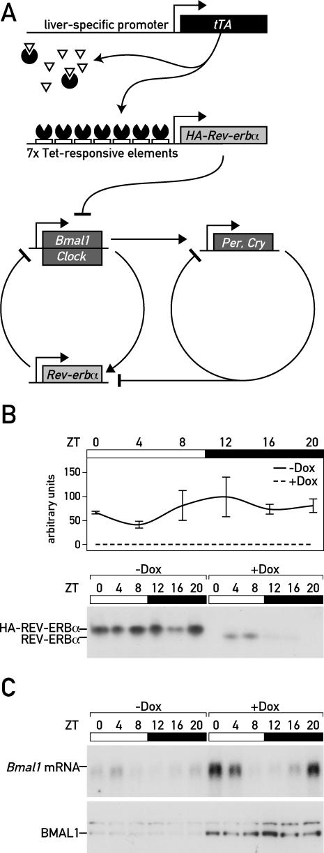
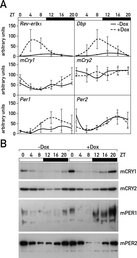
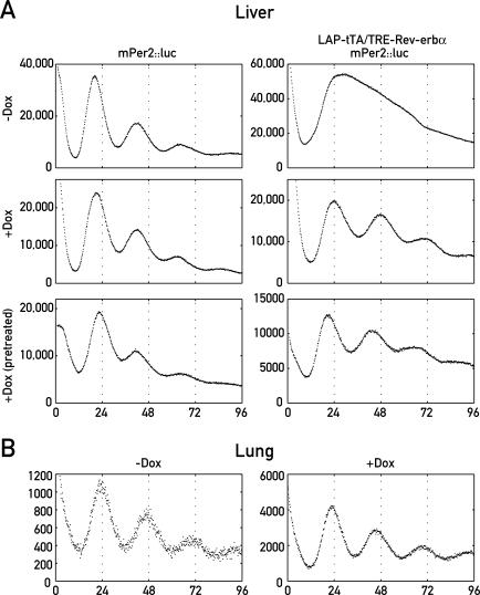
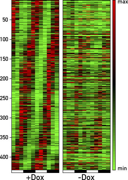
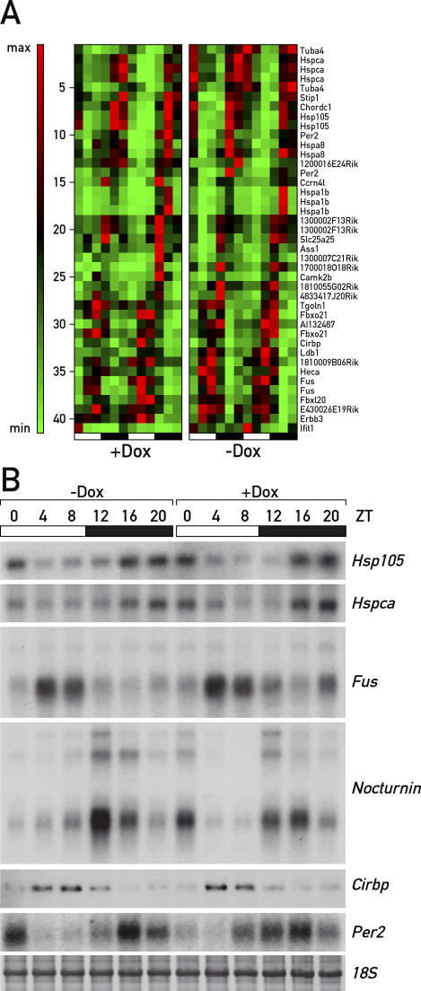
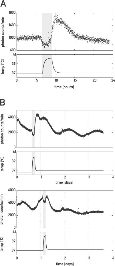
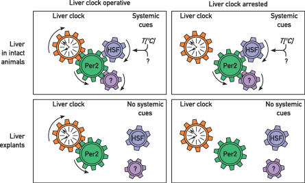
The mammalian circadian timing system consists of a master pacemaker in neurons of the suprachiasmatic nucleus (SCN) and clocks of a similar molecular makeup in most peripheral body cells. Peripheral oscillators are self-sustained and cell autonomous, but they have to be synchronized by the SCN to ensure phase coherence within the organism. In principle, the rhythmic expression of genes in peripheral organs could thus be driven not only by local oscillators, but also by circadian systemic signals. To discriminate between these mechanisms, we engineered a mouse strain with a conditionally active liver clock, in which REV-ERBalpha represses the transcription of the essential core clock gene Bmal1 in a doxycycline-dependent manner. We examined circadian liver gene expression genome-wide in mice in which hepatocyte oscillators were either running or arrested, and found that the rhythmic transcription of most genes depended on functional hepatocyte clocks. However, we discovered 31 genes, including the core clock gene mPer2, whose expression oscillated robustly irrespective of whether the liver clock was running or not. By contrast, in liver explants cultured in vitro, circadian cycles of mPer2::luciferase bioluminescence could only be observed when hepatocyte oscillators were operational. Hence, the circadian cycles observed in the liver of intact animals without functional hepatocyte oscillators were likely generated by systemic signals. The finding that rhythmic mPer2 expression can be driven by both systemic cues and local oscillators suggests a plausible mechanism for the phase entrainment of subsidiary clocks in peripheral organs.


Conflict of interest statement

Competing interests. The authors have declared that no competing interests exist.

Figures

Figure 1
Figure 1. Conditional Repression of Bmal1 Transcription in Hepatocytes
(A) Hepatocyte-specific, Dox-dependent expression of HA-REV-ERBα was achieved by placing a 5′-HA-tagged REV-ERBα cDNA transgene under the control of seven TREs (Tet-responsive elements). In the liver of mice expressing the tetracycline (Tet)-responsive transactivator (tTA) from the hepatocyte-specific C/ebpβ-LAP locus control region, HA-Rev-erbα transcription is constitutively repressed in the absence of the tetracycline analog Dox (tet-off system). This leads to an attenuation of circadian oscillator function, since Bmal1 is required for circadian rhythm generation. (B) LAP-tTA/TRE-Rev-erbα double transgenic mice display high HA-Rev-erbα mRNA and protein levels throughout the day in the absence of Dox (−Dox). In the presence 3 g/kg Dox in the food (+Dox), neither HA-Rev-erbα mRNA nor protein can be detected. (C) The levels of both Bmal1 mRNA and BMAL1 protein are dramatically down-regulated in the absence of Dox (compare the lanes on the left to those on the right).
Figure 2
Figure 2. The Expression of Clock and Clock-Controlled Genes Can Be Differentially Affected by HA-REV-ERBα Overexpression
(A) TaqMan real-time RT-PCR of cDNA was performed with liver whole-cell RNA for the transcripts of Dbp, endogenous Rev-erbα, mCry1, mCry2, mPer1, and mPer2 from untreated LAP-tTA/TRE-Rev-erbα mice (solid lines; −Dox) and Dox-treated LAP-tTA/TRE-Rev-erbα mice (dotted lines; +Dox). (B) Western blot analysis of liver nuclear extracts from LAP-tTA/TRE-Rev-erbα mice that were fed with normal chow (−Dox) or Dox-treated chow (+Dox). In accordance with the temporal mRNA profiles shown in (A) and (B), mCRY1 and mPER1 display reduced levels in untreated mice, whereas mPer2 and mCry2 accumulate to similar levels in nuclei of Dox-treated and untreated animals. The varying mPER1 migration is probably due to oscillating protein phosphorylation and dephosphorylation (see [18]).
Figure 3
Figure 3. Temporal Luminescence Profiles of Organ Explants from TRE-Rev-erbα/LAP-tTA/mPer2::luc Triple Transgenic Mice
(A) Liver slices from mPer2::luc (left) and LAP-tTA/TRE-Rev-erbα/mPer2::luc (right) mice were cultured in luciferin-containing medium in the absence (−Dox) or presence (+Dox) of 10-ng/μl Dox. Luminescence was recorded using photomultiplier tubes. −Dox and +Dox samples are from the same animal; +Dox (pretreated) samples are from mice that have received two intraperitoneal injections of Dox 48 h and 24 h before being sacrificed. (B) Lung explants from LAP-tTA/TRE-Rev-erbα/mPer2::luc mice were cultured as above in the presence or absence of Dox.
Figure 4
Figure 4. Phase Map of Circadian Transcripts Revealed by Genome-Wide Transcriptome Profiling
LAP-tTA/TRE-Rev-erbα mice fed with Dox-supplemented chow (+Dox) or normal chow (−Dox) were sacrificed at twelve 4-h intervals, and the liver transcriptomes were profiled by Affymetrix oligonucleotide microarray hybridization. Circadian transcripts were retrieved as outlined in Materials and Methods from the 24 datasets, and their temporal expression patterns were aligned according to phase. Note that the circadian accumulation of most transcripts is severely blunted in the livers of untreated mice. The heat scale to the right of the panels represents amplitudes in a linear scale, where green and red represent minimal and maximal expression levels, respectively.
Figure 5
Figure 5. Systemically Driven Circadian Genes Are Unaffected by REV-ERBα Overexpression
(A) A subset of transcripts whose circadian accumulation is not significantly affected by the HA-REV-ERBα overexpression is displayed according to the criteria used for (A). We conclude that the circadian rhythms of these genes are driven by systemic timing cues. (B) Northern blot quantification of some systemically driven circadian genes. Some of the results of (A) were validated by Northern blotting. For all six genes tested (Hsp105, Hspca, Fus, Nocturnin/Ccrn4l, Cirbp, and mPer2), neither amplitude nor phase is affected in a significant manner by REV-ERBα overexpression. The sharpness of the bands for 18S RNA and Cirbp are due to a shorter migration of this particular formaldehyde agarose gel.
Figure 6
Figure 6. Heat-Shock Induction of mPer2
(A) Liver explants of LAP-tTA/TRE-Rev-erbα/mPer2::luc mice were cultured as in Figure 3 and subjected to heat shock (150 min at ∼40 °C) using homemade culture-dish heating devices, and luminescence was recorded as in Figure 3. Temperature plots are extrapolated from periodic temperature measurement. The time window during which organ cultures were exposed to an elevated temperature is depicted by a grey box. (B) Lung explants were subjected to heat shock as in (A) at two different circadian times.
Figure 7
Figure 7. Model for the Synchronization of Liver Oscillators
In the intact animal, the phase of circadian mPer2 cycles is dictated by systemic Zeitgeber cues such as temperature or chemical cues influencing HSF activity (see text). Since mPER2 is also an integral part of the clockwork circuitry, this protein might confer the phase of systemic Zeitgebers to the local oscillator. If the oscillator is inactivated (e.g., by the repression of Bmal1), mPER2 is still expressed in a circadian manner in the intact animal. Under free-running conditions (i.e., in liver explants cultured in vitro), rhythmic mPer2 expression persists, but with the phase and period imposed by the local oscillators. However, when this oscillator is arrested, the expression of mPer2 and probably that of all clock and clock-controlled genes becomes arrhythmic.

Comment in

References

    1. Nagoshi E, Brown SA, Dibner C, Kornmann B, Schibler U. Circadian gene expression in cultured cells. Methods Enzymol. 2005;393:543–557. - PubMed
    1. Yoo S-H, Yamazaki S, Lowrey PL, Shimomura K, Ko CH, et al. PERIOD2::LUCIFERASE real-time reporting of circadian dynamics reveals persistent circadian oscillations in mouse peripheral tissues. Proc Natl Acad Sci U S A. 2004;101:5339–5346. - PMC - PubMed
    1. Welsh DK, Yoo SH, Liu AC, Takahashi JS, Kay SA. Bioluminescence imaging of individual fibroblasts reveals persistent, independently phased circadian rhythms of clock gene expression. Curr Biol. 2004;14:2289–2295. - PMC - PubMed
    1. Hannibal J. Neurotransmitters of the retino-hypothalamic tract. Cell Tissue Res. 2002;309:73–88. - PubMed
    1. Damiola F, Le Minh N, Preitner N, Kornmann B, Fleury-Olela F, et al. Restricted feeding uncouples circadian oscillators in peripheral tissues from the central pacemaker in the suprachiasmatic nucleus. Genes Dev. 2000;14:2950–2961. - PMC - PubMed

Publication types

MeSH terms