. 2007 May 15;404(1):131-40.
doi: 10.1042/BJ20061747.

The major determinant of the heparin binding of glial cell-line-derived neurotrophic factor is near the N-terminus and is dispensable for receptor binding

Affiliations

The major determinant of the heparin binding of glial cell-line-derived neurotrophic factor is near the N-terminus and is dispensable for receptor binding

Ivan Alfano et al. Biochem J. .

Abstract

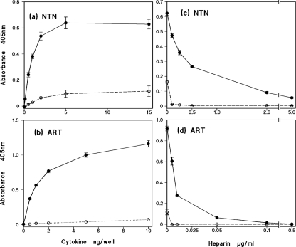
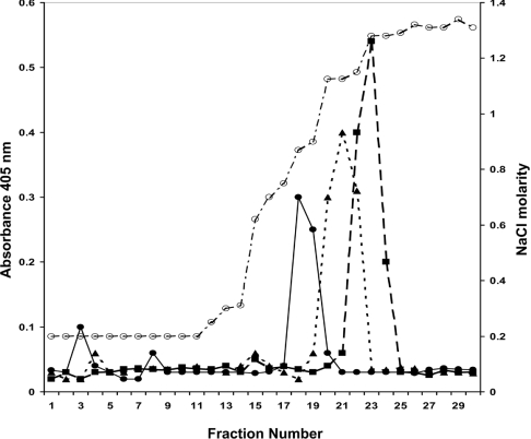
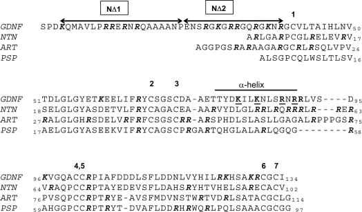
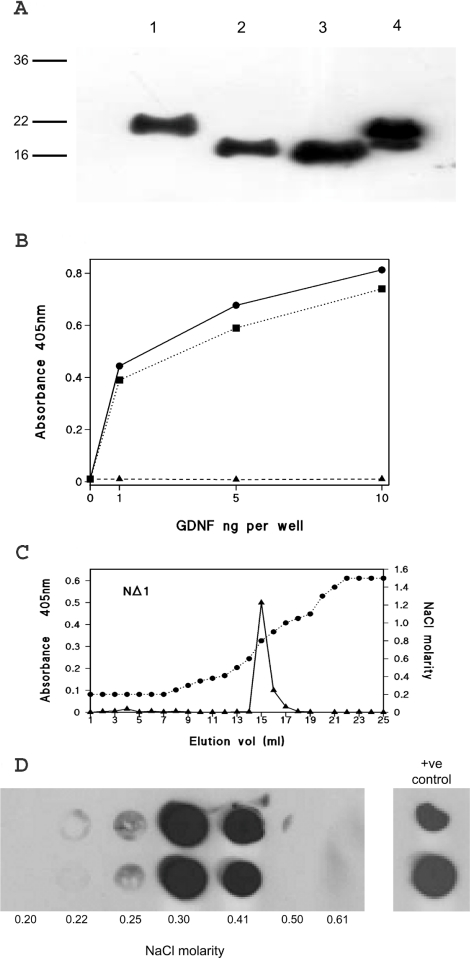
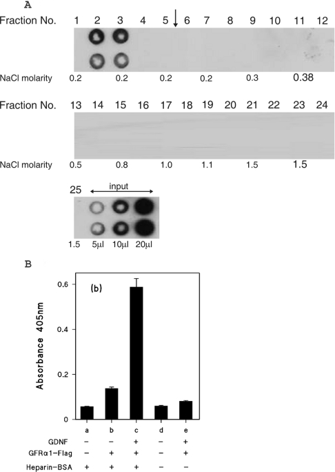
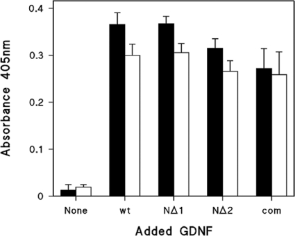
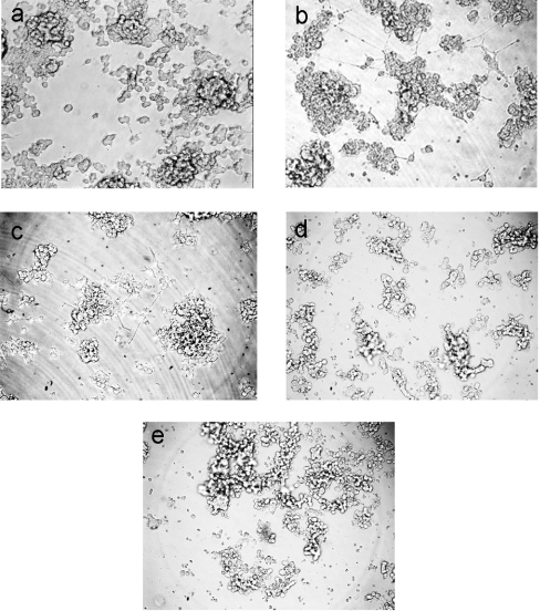
GDNF (glial cell-line-derived neurotrophic factor), and the closely related cytokines artemin and neurturin, bind strongly to heparin. Deletion of a basic amino-acid-rich sequence of 16 residues N-terminal to the first cysteine of the transforming growth factor beta domain of GDNF results in a marked reduction in heparin binding, whereas removal of a neighbouring sequence, and replacement of pairs of other basic residues with alanine had no effect. The heparin-binding sequence is quite distinct from the binding site for the high affinity GDNF polypeptide receptor, GFRalpha1 (GDNF family receptor alpha1), and heparin-bound GDNF is able to bind GFRalpha1 simultaneously. The heparin-binding sequence of GDNF is dispensable both for GFRalpha1 binding, and for activity for in vitro neurite outgrowth assay. Surprisingly, the observed inhibition of GDNF bioactivity with the wild-type protein in this assay was still found with the deletion mutant lacking the heparin-binding sequence. Heparin neither inhibits nor potentiates GDNF-GFRalpha1 interaction, and the extracellular domain of GFRalpha1 does not bind to heparin itself, precluding heparin cross-bridging of cytokine and receptor polypeptides. The role of heparin and heparan sulfate in GDNF signalling remains unclear, but the present study indicates that it does not occur in the first step of the pathway, namely GDNF-GFRalpha1 engagement.


Figures

Figure 1
Figure 1. ELISA of the binding of recombinant human NTN and ART to immobilized heparin
Dose-dependence of (a) NTN and (b) ART binding. Competitive binding after pre-incubation with increasing concentrations of soluble heparin of 2 ng/well NTN (c) and 8 ng/well ART (d). The solid lines and closed symbols represent wells coated with heparin–BSA complex; the open symbols and dotted lines indicate control wells coated with mock-conjugated BSA. The results shown are means±S.E.M. for a single representative experiment, and each data point taken is from four or five replicate wells.
Figure 2
Figure 2. Heparin affinity chromatography of rat GDNF, human ART and human NTN
Commercial recombinant GFLs (500 ng) dissolved in PBS were separately applied to a HiTrap Heparin column. Eluate fractions (1 ml) were assayed for GFL by heparin-binding ELISA. Salt concentrations (open circles, dotted and dashed line) were determined by conductivity measurements as shown on the right-hand axis, by standardizing with a range of solutions of known concentration dissolved in PBS. Fractions were assayed for GDNF (closed circles and solid line), NTN (closed triangles and dotted line), and ART (closed squares and dashed line) by heparin-binding ELISA (left-hand axis).
Figure 3
Figure 3. Alignment of the amino acid sequences of the GFLs
The sequences of the mature, secreted forms of rat GDNF, and human ART, NTN and PSN are shown. The human GDNF sequence differs from that of the rat by seven residues. The residues comprising the α-helix of GDNF are indicated. The residues deleted in the two truncation mutations NΔ1 and NΔ2 are shown, and the residues subjected to alanine replacement are underlined. Arginine and lysine residues are highlighted in bold italics.
Figure 4
Figure 4. Characterization of double point mutants of rat GDNF
(A) Western blot of baculovirus-expressed mutants of GDNF. Conditioned supernatants from infected Sf9 cells were subjected to ion-exchange chromatography on SP-Trisacryl and GDNF immunoreactivity fractions were dialysed against PBS. The loadings shown were adjusted to give comparable intensities of development. Lane 1, commercial rat GDNF; lane 3, baculovirus-expressed wild-type GDNF; lane 5, K81A/K84A double point mutant; and, lane 7, R88A/R90A double point mutant. Lanes 2, 4 and 6 show similarly processed culture supernatants from mock-infected Sf9 cells, treated and grown in parallel to the cultures yielding the supernatant shown on the immediate right-hand side. (B) Heparin chromatography of wild-type (Wt) and double point mutants of GDNF. Samples of the immunoreactive SP-Trisacryl eluate fractions were dialysed against PBS and applied to 1 ml HiTrap Heparin columns. Fractions were assayed for GDNF immunoreactivity by heparin-binding ELISA (closed squares and solid line) and for conductivity (closed circles and dashed line).
Figure 5
Figure 5. Characterization of partial N-terminal deleted mutants of GDNF
(A) Western blot of baculoviral-expressed mutants of GDNF following SP-Trisacryl ion-exchange chromatography. Lane 1, wild-type rat GDNF; lane 2, NΔ1-GDNF; lane 3, NΔ2-GDNF; and, lane 4, commercial mammalian cell expressed rat GDNF. (B) Heparin-binding ELISA of wild-type rat GDNF (closed circles and solid line), NΔ1 (closed squares and dotted line), and NΔ2 (closed triangles and dashed line). Each data point is the mean of triplicate wells. (C) and (D) Heparin affinity chromatography of GDNF N-terminal deletion mutants. (C) NΔ1; immunoreactivity detected by heparin-binding ELISA. (D) NΔ2; immunoreactivity detected by dot blotting. Duplicate dot blots of fractions from the beginning of the salt gradient are shown together with the molarity determined, this region of the elution being the only one showing immunoreactivity. Duplicate applications of the input are shown on the right-hand side.
Figure 6
Figure 6. Heparin affinity chromatography of GFRα1–FLAG chimaeric protein
(A) GFRα1–FLAG partially purified by ion-exchange chromatography was applied to a heparin Hi-trap column as described in the Experimental section for GDNF, except that the linear NaCl gradient was 0.18–2 M. The gradient was applied after fraction 5, as indicated by the vertical arrow. The NaCl molarities of the odd numbered fractions, as determined by conductivity measurements are shown. (B) Binding of GFRα1–FLAG to heparin-immobilized GDNF. GDNF (2.5 ng/well) was captured on wells coated with either heparin–BSA complex (columns a–c) or mock-conjugated BSA (columns d and e) as described for the heparin-binding ELISA. GFRα1–FLAG was then added to wells (columns b, c and e) at a quantity of 20 ng/well, as estimated by comparison with the GFRα1–Fc Western blotting. All wells were developed with anti-FLAG and alkaline phosphatase-labelled second antibody. The results are shown as means±S.E.M. for four replicate wells from a single representative experiment.
Figure 7
Figure 7. inding of wild-type GDNF and deletion mutants NΔ1 and NΔ2 to GFRα1 in the presence and absence of heparin
FRa1–FLAG was incubated with GDNF variants for 30 min in the absence (closed bars) or presence (open bars) of 2 μg/ml heparin, before capture on anti-FLAG coated wells and subsequent detection with anti-FLAG and secondary antibodies. The quantity of the GDNF variants used was judged by Western blotting to be comparable with 2 ng/well of commercial GDNF, a quantity giving high, but sub-maximal, absorbance. The absorbance shown was blanked against anti-FLAG coated wells with no GFRα1–FLAG or GDNF. Values are the means for triplicate wells and shown ± S.D. wt, wild-type GDNF; com; commercial recombinant rat GDNF.
Figure 8
Figure 8. Neurite outgrowth activity in vitro of wild-type GDNF and deletion mutant NΔ2
(a) No GDNF. (b) and (d) Wild-type GDNF. (c) and (e) NΔ2-GDNF. Incubations were performed in the absence (ac) or presence (d and e) of 1 μg/ml heparin. GFRα1–Fc (2 ng/well) was used throughout and, where present, the baculovirus expressed GDNF variants were employed at quantities judged by Western blotting to be equivalent to 10 ng/well.

References

    1. Baloh R., Enomoto H., Johnson E., Milbrandt J. The GDNF family ligands and receptors: implications for neural development. Curr. Opin. Neurobiol. 2000;10:103–110. - PubMed
    1. Carmillo P., Dago L., Day E., Worley D., Rossomando A., Walus L., Orozco O., Buckley C., Miller S., Tse A., et al. Glial cell line-derived neurotrophic factor (GDNF) receptor α-1 (GFRα1) is highly selective for GDNF versus artemin. Biochemistry. 2005;44:2545–2554. - PubMed
    1. Poteryaev D., Titievsky A., Sun Y. F., Thomas-Crusells J., Lindahl M., Billaud M., Arumae U., Saarma M. GDNF triggers a novel ret-independent Src kinase family-coupled signalling via a GPI-linked GDNF receptor α1. FEBS Lett. 1999;463:63–66. - PubMed
    1. Trupp M., Scott R., Whittlemore S. R., Ibanez C. F. Ret-dependent and -independent mechanisms of glial cell line-derived neurotrophic factor signalling in neuronal cells. J. Biol. Chem. 1999;274:20885–20894. - PubMed
    1. Popsueva A., Poteryaev D., Arighi E., Meng X., Angers-Loustau A., Kaplan D., Saarma M., Sariola H. GDNF promotes tubulogenesis of GFR1-expressing MDCK cells by Src-mediated phosphorylation of Met receptor protein kinase. J. Cell Biol. 2003;161:119–129. - PMC - PubMed

Publication types

MeSH terms

Substances