

doi: 10.1016/j.jneumeth.2007.01.004.
Epub 2007 Jan 13.
Micro-field evoked potentials recorded from the porcine sub-dural cortical surface utilizing a microelectrode array
Affiliations
- PMID: 17298849
- PMCID: PMC2223486
- DOI: 10.1016/j.jneumeth.2007.01.004
Item in Clipboard
Micro-field evoked potentials recorded from the porcine sub-dural cortical surface utilizing a microelectrode array
J Neurosci Methods.
.
Abstract
A sub-dural surface microelectrode array designed to detect micro-field evoked potentials has been developed. The device is comprised of an array of 350-microm square gold contacts, with bidirectional spacing of 150 microm, contained within a polyimide Kapton material. Cytotoxicity testing suggests that the device is suitable for use with animal and human patients. Implementation of the device in animal studies revealed that reliable evoked potentials could be acquired. Further work will be needed to determine how these micro-field potentials, which demonstrate selectivity for one eye, relate to the distribution of the ocular dominance columns of the occipital cortex.
Figures
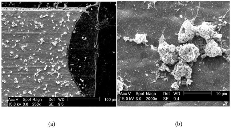
(a) SEM image of human cortical cells cultured on the Kapton and gold microelectrode surface (the light portion is a gold contact and the dark is the polyimide Kapton); (b) SEM image showing axonic and/or dendritic growth from the human cortical cells on the gold microelectrode contact surface.

Exposed sub-dural cortical surface of porcine occipital cortex. Microelectrode recordings were obtained from the left occipital cortex (area 17). Location of electrode placement is indicated.

Surface microfield potentials collected using the Kapton and gold microelectrode arrays. The individual electrode contact site and the corresponding collected potentials are shown. Right eye stimulation potentials plots are shown above left eye stimulation potential plots for each electrode contact. Each plot shows Voltage (V range = -15.0 to +15.0 mV) on the Y-axis and time (t range = 0 to +250.0 msec) on the X-axis. For each plot, the stimulus occurred at time = 0.

Averaged surface microfield potentials collected using the Kapton and gold microelectrode arrays. The individual electrode contact site and the corresponding collected potentials are shown. Right eye stimulation potentials plots are shown above left eye stimulation potential plots for each electrode contact. Each plot shows Voltage (V range = -15.0 to +15.0 mV) on the Y-axis and time (t range = 0 msec to +250.0 msec) on the X-axis. For each plot, the stimulus occurred at time = 0.

Averaged potentials collected using the penetrating electrodes for electrodes resoponding to the left eye (a) and right eye (b). For each plot, the stimulus occurred at time = 0.

(a) Schematic of a 32 contact gold and Kapton microelectrode array utilizing printed circuitry; (b) A gold and Kapton 32 contact microelectrode array comprised of gold contacts (200 micron squares with bidirectional pitch of 400 microns in a 4 by 8 grid pattern). (The Ohio State University and Valtronic, USA Inc. Patent pending.)
References
-
- Bernadette F. Stereotaxic atlas of the pig brain. Brain Res. Bull. 1999;49:1–138. - PubMed
-
- Black J. Biological Performance of Materials: Fundamentals of Biocompatibilty. Marcel Dekker, Inc; New York: 1999. pp. 323–425.
-
- Bugbee NM, Goldmann-Rakic P. Columnar organization of cortico-cortical projections in squirrel and rhesus monkey: similarity of column width in species differing in cortical volume. J. Comp. Neurol. 1983;220:355–64. - PubMed
-
- Eckhorn R, Stett A, Schanze T, Gekeler F, Schwahn H, Zrenner E, Wilms M, Eger M, Hesse L. Physiological functional evaluation of retinal implants in animal models. Ophthalmologe: Zeischrift der Deutschen Ophthalmologischen Gesellschaft. 2001;98:369–75. - PubMed
Publication types
MeSH terms
Grants and funding
LinkOut - more resources
Full Text Sources
Other Literature Sources
