. 2007 Feb 20;104(8):2649-54.
doi: 10.1073/pnas.0611503104. Epub 2007 Feb 13.

A natively unfolded yeast prion monomer adopts an ensemble of collapsed and rapidly fluctuating structures

Affiliations

A natively unfolded yeast prion monomer adopts an ensemble of collapsed and rapidly fluctuating structures

Samrat Mukhopadhyay et al. Proc Natl Acad Sci U S A. .

Abstract

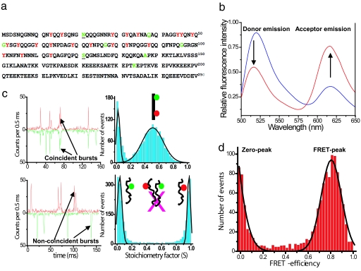
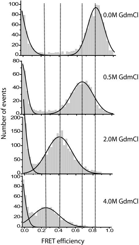
The yeast prion protein Sup35 is a translation termination factor, whose activity is modulated by sequestration into a self-perpetuating amyloid. The prion-determining domain, NM, consists of two distinct regions: an amyloidogenic N terminus domain (N) and a charged solubilizing middle region (M). To gain insight into prion conversion, we used single-molecule fluorescence resonance energy transfer (SM-FRET) and fluorescence correlation spectroscopy to investigate the structure and dynamics of monomeric NM. Low protein concentrations in these experiments prevented the formation of obligate on-pathway oligomers, allowing us to study early folding intermediates in isolation from higher-order species. SM-FRET experiments on a dual-labeled amyloid core variant (N21C/S121C, retaining wild-type prion behavior) indicated that the N region of NM adopts a collapsed form similar to "burst-phase" intermediates formed during the folding of many globular proteins, even though it lacks a typical hydrophobic core. The mean distance between residues 21 and 121 was approximately equal to 43 A. This increased with denaturant in a noncooperative fashion to approximately equal to 63 A, suggesting a multitude of interconverting species rather than a small number of discrete monomeric conformers. Fluorescence correlation spectroscopy analysis of singly labeled NM revealed fast conformational fluctuations on the 20- to 300-ns time scale. Quenching from proximal and distal tyrosines resulted in distinct fast and slower fluctuations. Our results indicate that native monomeric NM is composed of an ensemble of structures, having a collapsed and rapidly fluctuating N region juxtaposed with a more extended M region. The stability of such ensembles is likely to play a key role in prion conversion.


Conflict of interest statement

The authors declare no conflict of interest.

Figures

Fig. 1.
Fig. 1.
Amino acid sequence, ensemble, and single-molecule data for NM. (a) Sequence of the prion domain (NM) of Sup35 showing residues mutated to cysteine in green for single fluorescence labeling (Alexa Fluor 488). Underscores (21 and 121) indicate the dual cysteine mutation for donor (Alexa Fluor 488) and acceptor (Alexa Fluor 594) labeling for FRET studies. Tyrosines are shown in red. (b) Steady-state ensemble fluorescence spectra of double-labeled NM (0.1 μM) showing energy transfer under denatured (6 M GdmCl) conditions (blue) and under native condition (red) obtained by exciting the donor (λex 488 nm). (c) Two-color single-molecule fluorescence coincidence: Coincident bursts and stoichiometric factor histogram for dual-labeled DNA (non-FRET) standard sample (Upper) and for a mixture of two single-labeled NM (100 pM each) under native condition showing no intermolecular association under this condition (Lower). (d) Single-molecule FRET-efficiency histogram measured ratiometrically for NM under native conditions. Black curves are the best fits using Gaussian functions. See SI Text for details.
Fig. 2.
Fig. 2.
FRET-efficiency histograms of NM at various concentrations of GdmCl. Dotted lines are drawn to show the progressive shift in the FRET-peak from 0.8 to 0.3.
Fig. 3.
Fig. 3.
Progressive E-shifts and Gaussian simulations support fast dynamics. (a) Plot of FRET efficiency vs. denaturant concentration. The solid line is the exponential fit to guide the eye. (b) Expected FRET-efficiency distribution calculated by taking shot-noise into account for the two limiting cases of fast (solid line) and slow (dashed line) conformational averaging with respect to the observation time (0.5 ms) of a Gaussian chain (for mean E of 0.8). Observed histogram is shown in gray.
Fig. 4.
Fig. 4.
FCS autocorrelation of NM labeled with Alexa Fluor 488 at 21 (a) and 184 (b) under native condition showing the fluctuation and diffusion components. (Insets) The complete autocorrelation curves.

References

    1. Prusiner SB. Proc Natl Acad Sci USA. 1998;95:13363–13383. - PMC - PubMed
    1. Caughey B. Nat Med. 2000;6:751–754. - PubMed
    1. Chien P, Weissman JS. Nature. 2001;410:223–227. - PubMed
    1. Shorter J, Lindquist S. Nat Rev Genet. 2005;6:435–450. - PubMed
    1. Tuite MF, Cox BS. Nat Rev Mol Cell Biol. 2003;4:878–889. - PubMed

Publication types

MeSH terms

LinkOut - more resources