. 2007 Feb 20;104(8):2861-6.
doi: 10.1073/pnas.0611487104. Epub 2007 Feb 13.

The Fas pathway is involved in pancreatic beta cell secretory function

Affiliations

The Fas pathway is involved in pancreatic beta cell secretory function

Desiree M Schumann et al. Proc Natl Acad Sci U S A. .

Abstract

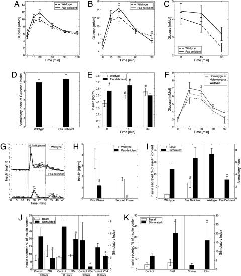
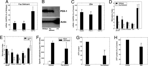
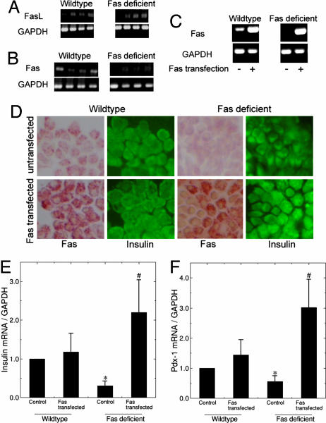
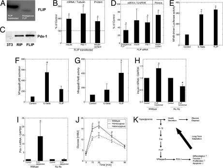
Pancreatic beta cell mass and function increase in conditions of enhanced insulin demand such as obesity. Failure to adapt leads to diabetes. The molecular mechanisms controlling this adaptive process are unclear. Fas is a death receptor involved in beta cell apoptosis or proliferation, depending on the activity of the caspase-8 inhibitor FLIP. Here we show that the Fas pathway also regulates beta cell secretory function. We observed impaired glucose tolerance in Fas-deficient mice due to a delayed and decreased insulin secretory pattern. Expression of PDX-1, a beta cell-specific transcription factor regulating insulin gene expression and mitochondrial metabolism, was decreased in Fas-deficient beta cells. As a consequence, insulin and ATP production were severely reduced and only partly compensated for by increased beta cell mass. Up-regulation of FLIP enhanced NF-kappaB activity via NF-kappaB-inducing kinase and RelB. This led to increased PDX-1 and insulin production independent of changes in cell turnover. The results support a previously undescribed role for the Fas pathway in regulating insulin production and release.


Conflict of interest statement

The authors declare no conflict of interest.

Figures

Fig. 1.
Fig. 1.
Fas regulates β cell secretory function. Blood glucose levels after i.p. injection of 2 mg of glucose (A) and 1 mg of glucose (B) per gram of body weight in male Fas-deficient B6.MRLlpr and wild-type C57BL/6j mice aged 7–8 weeks. ∗, P < 0.05, Fas-deficient versus wild type (n = 15 and 10 for each group in A and B, respectively). (C) Blood glucose levels after i.p. injection of 0.5 units of insulin per kilogram of body weight in male Fas-deficient and wild-type mice. ∗, P < 0.05, Fas-deficient versus wild type (n = 15 for each group). (D) Insulin-stimulated 2-deoxyglucose uptake in adipocytes isolated from Fas-deficient and wild-type mice (n = 2, each in hexaplicate). (E) Insulin levels after i.p. injection of 2 mg of glucose per gram of body weight in male Fas-deficient and wild-type mice (n = 15 for each group). ∗, P < 0.05 relative to wild type at time point 0 min; #, P < 0.05 relative to wild type at time point 15 min. (F) Blood glucose levels after i.p. injection of glucose in male prediabetic NODβFas−/− (homozygous), NODβFas+/− (heterozygous), and NODβFas+/+ (wild-type) littermate mice aged 5–6 weeks. ∗, P < 0.05 (n = 5–10 for each group). (G and H) Glucose-induced insulin secretion in perfused pancreata from Fas-deficient and wild-type mice. Pancreata were perfused with basal solution (2.8 mM glucose) for 30 min, and then glucose was increased to 16.7 mM for the indicated period (n = 3). (I) Percentage of islet insulin content released during a 1-h incubation at 3.3 mM (basal) and 16.7 mM (stimulated) glucose after an 8-day culture period of islets isolated from Fas-deficient and wild-type mice (Left) and the corresponding stimulatory index of insulin secretion (Right) (n = 4, each in hexaplicate). Insulin content was 0.12 ± 0.02 ng/ml for wild-type mice and 0.14 ± 0.03 ng/ml for Fas-deficient mice. ∗, P < 0.001. (J) Percentage of islet insulin content released during successive 1-h incubations at 3.3 mM (basal) and 16.7 mM (stimulated) glucose after 6-h and 4-day incubation periods of islets isolated from human pancreata in the presence of 500 ng/ml isotype IgG (control) or 500 ng/ml antagonistic anti-Fas antibody (ZB4) (Left) and the corresponding stimulatory index (Right) (n = 3, each in triplicate). Insulin content for control islets was 0.04 ± 0.01 and 0.03 ± 0.002 ng/ml, and insulin content for ZB4-treated islets was 0.05 ± 0.01 and 0.03 ± 0.01 ng/ml after 6 h and 4 days, respectively. ∗, P < 0.001. (K) Percentage of islet insulin content released during successive 1-h incubations at basal and stimulated glucose after incubation of human islets in the presence of FasL for 48 h. Insulin content for control was 0.04 ± 0.01 ng/ml, and insulin content for FasL-treated islets was 0.06 ± 0.01 ng/ml (n = 2, each done in quintuplicate).
Fig. 2.
Fig. 2.
Fas-deficient islets have decreased insulin and PDX1 expression and mitochondrial metabolism. (A) RT-PCR detection of insulin, PDX-1, and uncoupling protein 2 (UCP-2) mRNA expression in Fas-deficient and wild-type islets (n = 5 for each group). ∗, P < 0.05. (B) Immunoblotting of PDX-1 and actin of Fas-deficient and wild-type islets and from the 3T3 cell line (negative control). One of two experiments is shown. (C) RT-PCR detection of insulin, PDX-1, and uncoupling protein 2 (UCP-2) in human islets cultured for 4 days with 500 ng/ml isotype IgG (control) or 500 ng/ml antagonistic anti-Fas antibody (ZB4) (n = 3, each in duplicate). (D and E) Stimulatory index of insulin secretion during successive 1-h incubation at 3.3 mM (basal) and 16.7 mM (stimulated) glucose, 20 mM leucine, 10 mM α-ketoglutarate dimethyl ester (α-Ketoester), 10 mM succinic acid dimethyl ester (SAD), or 10 μM forskolin and 100 μM 3-isobutyl-1-methylxanthine (F/I), after a 4-day culture period of islets isolated from Fas-deficient and wild-type mice (D) and after a 4-day culture period of human islets in the presence of 500 ng/ml isotype IgG (control) or 500 ng/ml antagonistic anti-Fas antibody (ZB4) (E) (n = 3 from three different isolations/donors, each in triplicate). ∗, P < 0.05 vs. wild type or controls. (F) Islets isolated from wild-type and Fas-deficient mice were radiolabeled (25 min) with [3H]leucine in 2.8 mM (basal) and 16.7 mM (stimulated) glucose. Radiolabeled proinsulin plus insulin is presented as specifically immunoprecipitable radioactivity as a function of islet number (cpm per 100 islets) (n = 3). (G) After 1 day in culture, islets isolated from Fas-deficient and wild-type mice were incubated successively for 30 min in 2.8 mM glucose followed by an additional 10 min in 16.7 mM glucose and analyzed for stimulated ATP content per islet. ∗, P < 0.001 (n = 3, each in triplicate). (H) After a 4-day culture period, human islets cultured in the presence of 500 ng/ml isotype IgG (control) or 500 ng/ml antagonistic anti-Fas antibody (ZB4) were incubated successively for 30 min in 2.8 mM glucose followed by an additional 10 min in 16.7 mM glucose and analyzed for stimulated ATP content per islet. ∗, P < 0.05. Data are means of percentage relative to control for islets from three different donors, each plated in triplicate.
Fig. 3.
Fig. 3.
Fas and FasL are expressed in islets and regulate insulin and PDX-1 mRNA expression. (A and B) Shown is RT-PCR analysis of FasL (A) and Fas (B) expression in wild-type and Fas-deficient islets. Each lane represents an individual animal. GAPDH was used as control. (C–F) Fas-deficient and wild-type islets were transfected with a vector encoding for Fas and incubated for 8 days. (C) RT-PCR of Fas and GAPDH (control) expression in Fas-deficient and wild-type islets. (D) Immunostaining for Fas and insulin. (E and F) Quantitative RT-PCR detection of insulin (E) and PDX-1 (F) mRNA expression normalized to GAPDH (n = 3 for each group). ∗, P < 0.05 relative to wild type; #, P < 0.05 relative to Fas-deficient control.
Fig. 4.
Fig. 4.
FLIP regulates insulin and PDX-1 mRNA expression via the alternative pathway of NF-κB activation. INS-1E (A and E–G), wild-type, and aly/aly (B–D, H, and I) mouse islet cells were transfected with a mock vector or a vector encoding for FLIP (FLIP transfected) or exposed to siRNA to FLIP. (A) Immunoblotting of FLIP. (B and D) RT-PCR detection of insulin, PDX-1, and FLIP mRNA expression and islet insulin content. The level of mRNA expression was normalized to tubulin or GAPDH, and the results are expressed as mRNA levels relative to controls. (C) Immunoblotting of PDX-1 in islets and in 3T3 cells (negative control) transfected with the RIP vector alone and with RIP-FLIP. One of two experiments is shown. (E–G) Analysis of NF-κB activity by detection of luciferase activity after transfection of an NF-κB-driven firefly luciferase construct normalized to a cotransfected constitutive Renilla luciferase construct (E) and by detection of p50–p65 (F) and RelB (G) binding to an NF-κB consensus site. The effect of FLIP was compared with 150 pg/ml IL-1β (n = 4, each in duplicate). (H and I) RT-PCR of insulin (H) and PDX-1 (I) mRNA normalized to GAPDH (n = 3–4). ∗, P < 0.05 relative to wild-type control; #, P < 0.05 relative to aly/aly control. (J) Blood glucose levels after i.p. injection of glucose (2 mg/g of body weight) in male homozygous and heterozygous aly/aly mice and C57BL/6 (wild-type) mice. ∗, P < 0.05 homozygous, heterozygous vs. wild type (n = 6, 5, and 5, respectively, for each group). (K) Hypothetical model illustrating the consequence of limited hyperglycemia on β cell production of IL-1β in parallel with insulin secretion. The paracrine effect of IL-1β induces Fas engagement, which, in the presence of FLIP, leads to β cell proliferation, differentiation, and increased function.

References

    1. Rhodes CJ. Science. 2005;307:380–384. - PubMed
    1. Donath MY, Halban PA. Diabetologia. 2004;47:581–589. - PubMed
    1. Maedler K, Spinas GA, Lehmann R, Sergeev P, Weber M, Fontana A, Kaiser N, Donath MY. Diabetes. 2001;50:1683–1690. - PubMed
    1. Donath MY, Storling J, Maedler K, Mandrup-Poulsen T. J Mol Med. 2003;81:455–470. - PubMed
    1. Nolsoe RL, Hamid YH, Pociot F, Paulsen S, Andersen KM, Borch-Johnsen K, Drivsholm T, Hansen T, Pedersen O, Mandrup-Poulsen T. Genes Immun. 2006;7:316–321. - PubMed

Publication types

MeSH terms