. 2007 Feb 20;104(8):2797-802.
doi: 10.1073/pnas.0611259104. Epub 2007 Feb 13.

The Saccharomyces cerevisiae 14-3-3 proteins Bmh1 and Bmh2 directly influence the DNA damage-dependent functions of Rad53

Affiliations

The Saccharomyces cerevisiae 14-3-3 proteins Bmh1 and Bmh2 directly influence the DNA damage-dependent functions of Rad53

Takehiko Usui et al. Proc Natl Acad Sci U S A. .

Abstract

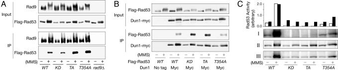
In this study, we mutated autophosphorylation sites in Rad53 based on their conservation with previously identified autophosphorylation sites in the mammalian Rad53 ortholog, Chk2. As with wild-type Rad53, the autophosphorylation mutant, rad53-TA, undergoes Mec1/Tel1-dependent interactions with Rad9 and Dun1 in response to genotoxic stress. Whereas rad53-TA in vitro kinase activity is severely impaired, the rad53-TA strains are not completely deficient for cell-cycle checkpoint functions, indicating that the mutant kinase retains a basal level of function. We describe a genetic interaction among Rad53, Dun1, and the 14-3-3 proteins Bmh1 and Bmh2 and present evidence that 14-3-3 proteins directly facilitate Rad53 function in vivo. The data presented account for the previously observed checkpoint defects associated with 14-3-3 mutants in Saccharomyces pombe and Saccharomyces cerevisiae. The 14-3-3 functional interaction appears to modulate Rad53 activity, reminiscent of 14-3-3's effect on human Raf1 kinase and distinct from the indirect mode of regulation by 14-3-3 observed for Chk1 or Cdc25.


Conflict of interest statement

The authors declare no conflict of interest.

Figures

Fig. 1.
Fig. 1.
Examination of rad53-T354A and rad53-TA proteins. (A and B) Western blot (WB) analyses of input cell extracts and Rad9 (A) and Myc (B) IP's from the indicated strains. Asynchronous cells (−) and 90 min after 0.03% MMS treatment (+) were examined. (C) Rad53 kinase activity was examined in the indicated strains. GST-dun1-KD was used as a substrate. (i) 32P-Flag-Rad53, (ii) Flag (Rad53) WB, (iii) 32P-GST-dun1-KD, and quantifications of Rad53 auto-(open bars) and transphosphorylation (filled bars) activity are shown.
Fig. 2.
Fig. 2.
Phenotypic analyses of rad53-T354A and rad53-TA mutants. HU sensitivity (A), MMS sensitivity (B), S/M (C), and G2/M cell cycle checkpoints (D) of the indicated strains were examined. Error bars represent standard deviation. (A) Cells arrested at G1 by α-factor were released into 200 mM HU and accessed for viability. (B) Asynchronous cells were treated with the indicated concentration of MMS for 20 min and tested for viability. (C) Percentage of the indicated cells that showed elongated spindle was determined by tubulin staining when treated with HU for 3 h as in A. The numbers in parentheses represent the actual observed numbers of cells with elongated spindle. (D) The indicated cdc13 cdc15 strains were arrested at G1 and released at the restrictive temperature (37°C) to examine the cdc13-induced G2/M DNA damage checkpoint. Nuclear division was monitored by DAPI staining (>200 cells counted). The y axis represented the percentage of cells that arrested at telophase (post-M).
Fig. 3.
Fig. 3.
BMH1/2 are high-copy suppressors of rad53-TA. (A) Growth of the rad53-TA, rad53-KD and rad53Δ transformed with single-copy RAD53, high-copy BMH2, and empty plasmids on 0 mM and 20 mM HU plates was assessed. Tenfold serial dilutions were plated. (B and C) Growth of rad53–21 (B), rad53-T354A and rad53-TA (C) transformed with the indicated plasmids on 20 mM or 40 mM HU plates was tested as in A.
Fig. 4.
Fig. 4.
Genetic requirement for BMH2 suppression. (A–C) The indicated rad53-TA strains transformed with the high-copy BMH2 (squares) and empty (triangles) vectors were incubated in 20 mM HU for 0, 3, and 6 h and accessed for viability. The dashed lines were RAD53, BMH2, and empty plasmid transformants of rad53-TA (A) and rad53-TA sml1Δ (B and C) from top. Note that sml1Δ suppresses lethality of the indicated genotypes in (B and C) but does not affect HU sensitivity of rad53-TA. (A) mrc1Δ rad53-TA (black symbols), and rad9Δ rad53-TA (gray symbols). (C) dun1Δ rad53-TA sml1Δ (open symbols) and dun1-fha rad53-TA sml1Δ (filled symbols). (D) G2/M DNA damage checkpoint was assessed in rad53-TA or dun1Δ rad53-TA sml1Δ cdc13 cdc15 transformed with the indicated plasmids as in Fig. 2D.
Fig. 5.
Fig. 5.
Co-IP of Rad53 and Bmh2. WB analyses of input cell extracts (i–iii) and Bmh2 IP (iv and v) from the indicated strains. WBs of Flag (Rad53) (i and iv), tubulin (ii) and Bmh1/2 (iii and v) were shown. PI, preimmune.
Fig. 6.
Fig. 6.
Analyses of phosphopeptide-binding site bmh2 mutants. (A) HU sensitivity of rad53-TA transformed with the high-copy BMH2 and indicated bmh2 mutant plasmids were tested. (B) HU and MMS sensitivity of the indicated genomic bmh2 mutants. (C) G2/M checkpoint of the indicated genotypes was examined as in Fig. 2D. bmh2-K51L and K51R are in bmh1Δ. (D) WB analyses of input cell extracts and Bmh2 IP's from the wild-type and indicated bmh2 mutant cells in bmh1Δ.

Similar articles

Cited by

References

    1. Zhou BB, Elledge SJ. Nature. 2000;408:433–439. - PubMed
    1. Nyberg KA, Michelson RJ, Putnam CW, Weinert TA. Annu Rev Genet. 2002;36:617–656. - PubMed
    1. Durocher D, Henckel J, Fersht AR, Jackson SP. Mol Cell. 1999;4:387–394. - PubMed
    1. Manke IA, Lowery DM, Nguyen A, Yaffe MB. Science. 2003;302:636–639. - PubMed
    1. Sanchez Y, Desany BA, Jones WJ, Liu Q, Wang B, Elledge SJ. Science. 1996;271:357–360. - PubMed

Publication types