. 2007 Feb 20;104(8):2885-90.
doi: 10.1073/pnas.0609359104. Epub 2007 Feb 13.

Evolutionary genomics reveals conserved structural determinants of signaling and adaptation in microbial chemoreceptors

Affiliations

Evolutionary genomics reveals conserved structural determinants of signaling and adaptation in microbial chemoreceptors

Roger P Alexander et al. Proc Natl Acad Sci U S A. .

Abstract

As an important model for transmembrane signaling, methyl-accepting chemotaxis proteins (MCPs) have been extensively studied by using genetic, biochemical, and structural techniques. However, details of the molecular mechanism of signaling are still not well understood. The availability of genomic information for hundreds of species enables the identification of features in protein sequences that are conserved over long evolutionary distances and thus are critically important for function. We carried out a large-scale comparative genomic analysis of the MCP signaling and adaptation domain family and identified features that appear to be critical for receptor structure and function. Based on domain length and sequence conservation, we identified seven major MCP classes and three distinct structural regions within the cytoplasmic domain: signaling, methylation, and flexible bundle subdomains. The flexible bundle subdomain, not previously recognized in MCPs, is a conserved element that appears to be important for signal transduction. Remarkably, the N- and C-terminal helical arms of the cytoplasmic domain maintain symmetry in length and register despite dramatic variation, from 24 to 64 7-aa heptads in overall domain length. Loss of symmetry is observed in some MCPs, where it is concomitant with specific changes in the sensory module. Each major MCP class has a distinct pattern of predicted methylation sites that is well supported by experimental data. Our findings indicate that signaling and adaptation functions within the MCP cytoplasmic domain are tightly coupled, and that their coevolution has contributed to the significant diversity in chemotaxis mechanisms among different organisms.


Conflict of interest statement

The authors declare no conflict of interest.

Figures
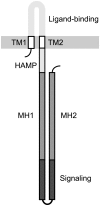
Fig. 1.
Fig. 1.
Schematic representation of the most common MCP topology and domain organization. Two transmembrane regions (TM1 and TM2) anchor the receptor in the membrane (shaded in gray). The sensory (ligand-binding) domain is extracellular, whereas the linker HAMP domain and the domain comprising two methylation regions (MH1 and MH2) and the signaling subdomain are in the cytoplasm. The boundaries of MH1, MH2, and the signaling subdomain are not well defined.
Fig. 2.
Fig. 2.
Schematic representation of MCP_CD features revealed by the multiple sequence alignment. The complete alignment is shown in SI Dataset 1. (A) Subdomain structure of major domain classes. The three subdomains, methylation helices, flexible bundle, and signaling, are indicated by medium, light, and dark gray, respectively. Each rectangle represents a group of two heptads. Heptads are numbered from N22 at the N terminus down to N01 at the center and then up from C01 at the center to C22 at the C terminus. This naming convention has the advantage that N- and C-terminal heptads with the same number are adjacent in the structure. Gap locations are shown in white. MCP classes are named 24H through 44H indicating the number of heptads (H). Experimentally determined methylation sites in class 36H MCPs from E. coli (28) and class 44H MCPs from B. subtilis (33, 40), H. salinarum (41, 42), and T. maritima (29, 43) are indicated by black circles. (B) Amino acid conservation within the MCP_CD. Position of each of 309 residues in the alignment is indicated by black columns. The column height shows the conservation level (see SI Text for details). Positions of the 44 seven-residue heptads (N22–C22) are indicated by background gray shading. The first and fourth (a and d) residues of each heptad are strongly conserved.
Fig. 3.
Fig. 3.
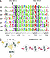
Family- and class-specific conservation in the signaling subdomain. (A) Sequence conservation in the seven major MCP classes. Two representative interdimer (N03b) and intradimer (C02d) interaction sites are indicated by arrows. (B) Visualization of selected inter- and intradimer sites in the E. coli Tsr trimer of dimers and T. maritima TM1143 hedgerow of dimers. Interdimer, N03b: Phe (Tsr), Glu (TM1143); intradimer, C02d: Val (both structures). Residue coloring: small (ASTG), green; hydrophobic (ILMV), black; aromatic (HFWY), yellow; negative (DE), red; polar (NQ), magenta; positive (KR), blue; and special (CP), cyan. MCP dimers are outlined in gray to highlight their different organization in the two structures.
Fig. 4.
Fig. 4.
Flexible bundle subdomain. (A) Representative knob (black) into hole (gray) packing in the E. coli Tsr protein. Residue numbers and heptad registers are shown. (B) Schematic representation of the arrangement of coiled-coil heptads in a four-helical bundle dimer of the MCP cytoplasmic domain, viewed axially from the top. Monomers A and B each have N-terminal (AN, BN) and C-terminal (AC, BC) helices joined by a hairpin loop at the base. Heptad registers a and d form a square-shaped knob layer at the core of the bundle. (C) A skewed knob layer of the coiled coil. Layers formed from large (black) and small (white) knobs are skewed from square- to diamond-shaped. (D) Stable and unstable knob layers. Stacks of knob layers with opposing skew are stable, whereas stacks of knob layers skewed in the same direction are unstable. (E) Lack of a classical coiled coil in the flexible bundle subdomain. Structures of the Tsr (Left) and TM1143 (Right) cytoplasmic domain show coiled-coil regions determined by the SOCKET algorithm with a 7.8-Å cutoff. Thin ribbons indicate regions where coiled coils were not detected. The bar indicates boundaries of the flexible bundle subdomain defined from the multiple sequence alignment. (F) Potential instability of the flexible bundle subdomain indicated by temperature factor. Colors indicate average temperature factor as follows. (i) Flexible bundle subdomain: “tendon” helices, red, very high; “bone” helices, orange, high; (ii) methylation subdomain, light blue, medium; (iii) signaling subdomain, dark blue, low. Detailed information is provided in SI Table 3. The bar indicates boundaries of the flexible bundle subdomain defined from the multiple sequence alignment.
Fig. 5.
Fig. 5.
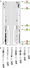
Methylation sites are conserved and located at class-specific positions. (A) Dot plot of the MCP_CD alignment showing conserved predicted methylation sites. A total of 1,656 sequences are shown. Heptads (N22–C22) are indicated by alternating gray shading. Positions of methylation sites matching the global consensus sequence are shown in black. Gaps are shown in white. Sequence logos of the methylation consensus sequence for each class are shown in color as in Fig. 3. (B) Homology models of major MCP_CD classes constructed from the T. maritima TM1143 structure (class 44H) show positions of the most common methylation sites (dark gray spheres) in each class. The signaling subdomain is shown in dark thick ribbons, the flexible bundle subdomain in light thin ribbons, and the methylation subdomain in dark thin ribbons. The glycine hinge is shown as a light gray sphere.

Comment in

  • Ancient chemoreceptors retain their flexibility.
    Parkinson JS. Parkinson JS. Proc Natl Acad Sci U S A. 2007 Feb 20;104(8):2559-60. doi: 10.1073/pnas.0700278104. Epub 2007 Feb 14. Proc Natl Acad Sci U S A. 2007. PMID: 17301220 Free PMC article. No abstract available.

References

    1. Segall JE, Block SM, Berg HC. Proc Natl Acad Sci USA. 1986;83:8987–8991. - PMC - PubMed
    1. Sourjik V, Berg HC. Proc Natl Acad Sci USA. 2002;99:123–127. - PMC - PubMed
    1. Alon U, Surette MG, Barkai N, Leibler S. Nature. 1999;397:168–171. - PubMed
    1. Endres RG, Wingreen NS. Proc Natl Acad Sci USA. 2006;103:13040–13044. - PMC - PubMed
    1. Francis NR, Wolanin PM, Stock JB, DeRosier DJ, Thomas DR. Proc Natl Acad Sci USA. 2004;101:17480–17485. - PMC - PubMed

Publication types

Substances

LinkOut - more resources