. 2007 Feb 20;104(8):2903-8.
doi: 10.1073/pnas.0608374104. Epub 2007 Feb 13.

Herpes simplex virus type 1 mediates fusion through a hemifusion intermediate by sequential activity of glycoproteins D, H, L, and B

Affiliations

Herpes simplex virus type 1 mediates fusion through a hemifusion intermediate by sequential activity of glycoproteins D, H, L, and B

Ravi P Subramanian et al. Proc Natl Acad Sci U S A. .

Abstract

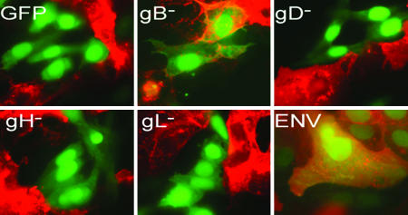
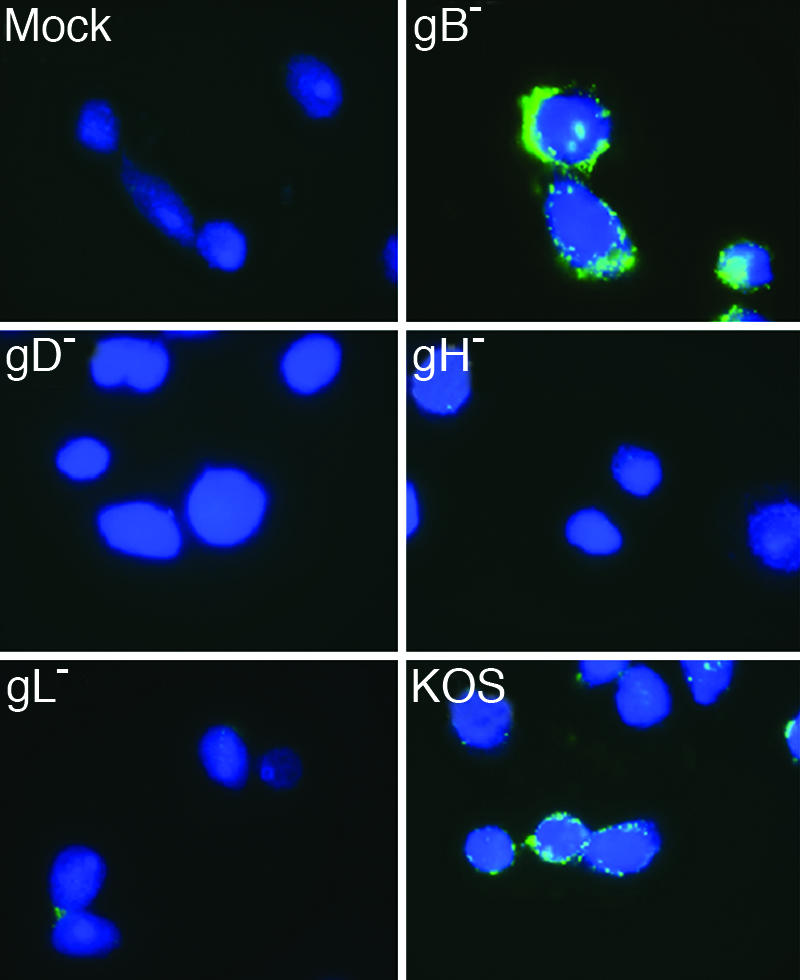
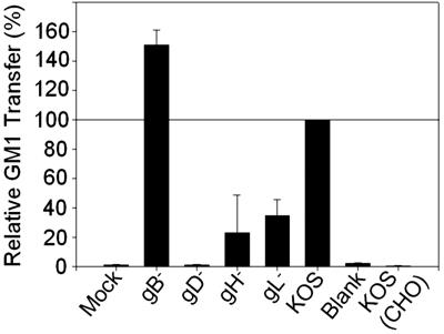
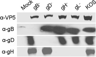
Virus-induced membrane fusion can be subdivided into three phases defined by studies of class I and class II fusion proteins. During Phase I, two membranes are brought into close apposition. Phase II marks the mixing of the outer membrane leaflets leading to formation of a hemifusion intermediate. A fusion pore stably forms and expands in Phase III, thereby completing the fusion process. Herpes simplex virus type 1 (HSV-1) requires four glycoproteins to complete membrane fusion, but none has been defined as class I or II. Therefore, we investigated whether HSV-1-induced membrane fusion occurred following the same general phases as those described for class I and II proteins. In this study we demonstrate that glycoprotein D (gD) and the glycoprotein H and glycoprotein L complex (gHL) mediated lipid mixing indicative of hemifusion. However, content mixing and full fusion required glycoprotein B (gB) to be present along with gD and gHL. Our results indicate that, like class I and II fusion proteins, fusion mediated by HSV-1 glycoproteins occurred through a hemifusion intermediate. In addition, both gB and gHL are probably directly involved in the fusion process. From this, we propose a sequential model for fusion via HSV-1 glycoproteins whereby gD is required for Phase I, gHL is required for Phase II, and gB is required for Phase III. We further propose that glycoprotein H and gB are likely to function sequentially to promote membrane fusion in other herpesviruses such as Epstein-Barr virus and human herpesvirus 8.


Conflict of interest statement

The authors declare no conflict of interest.

Figures

Fig. 1.
Fig. 1.
Glycoproteins necessary for hemifusion and complete fusion in the cell hemifusion assay. CHO cells were transfected with plasmids expressing GFP and all four fusion glycoproteins (ENV: gB, gD, gH, and gL) or three of the four (gB, gD, gH, and gL) glycoproteins. Transfected cells were overlaid with Vero-BG20 cells, fixed, and stained with TRITC-conjugated CTX (CTX-555) to detect GM1. Images were taken at ×100 magnification on a Zeiss Axiovert microscope. The experiment was performed three independent times, and images are from one representative experiment.
Fig. 2.
Fig. 2.
Glycoproteins required for GM1 transfer from virus to cell in virus fusion assay. Celltracker blue-labeled HveC-1 cells were inoculated with supernatants as depicted. The cells were treated with citrate buffer, fixed, and stained with FITC-CTX. Images were taken at ×100 magnification on a Zeiss Axiovert microscope. The experiment was performed three independent times, and images are from one representative experiment.
Fig. 3.
Fig. 3.
Relative GM1 transfer of HSV-1 and mutants viruses. HveC-1 cells were inoculated with culture medium (Blank) or supernatants from virus preparations. Cells were treated with citrate buffer, removed with Accutase, fixed, and stained with FITC-CTX to detect GM1 transfer. CHO-K1 cells were inoculated with KOS as a negative control [KOS (CHO)]. Results shown are the percentage of GM1+ relative to KOS GM1+ values. KOS GM1+ values ranged from 65% to 85% GM1+ cells of 30,000 total cells. Error bars represent the standard deviation of an average of two duplicate independent experiments.
Fig. 4.
Fig. 4.
Virus quantification. Equal volumes of concentrated virus supernatants were subjected to SDS/PAGE on an 8–16% gradient gel, transferred to nitrocellulose, and incubated with anti-VP5, anti-gD, anti-gH, or anti-gB. Blots were visualized as described in Materials and Methods. Quantitation was performed by using VP5 for each virus preparation made. Results are representative of those conducted on eight different sets of virus preparations. Anti-gB, anti-gD, and anti-gH blots are representative blots taken from one of three separate virus preparations.
Fig. 5.
Fig. 5.
Ab blocking of lipid transfer from virus to cells. Antibody (100 μg) or no antibody was added to ≈1 × 10⁷ pfu of concentrated gB or KOS virus supernatant for 1 h, followed by addition of supernatants to Celltracker red-labeled HveC-1 cells as described previously. (A) KOS, no Ab. (B) KOS, 100 μg of 52S. (C) KOS, 100 μg of H1817. (D) gB, no Ab. (E) gB, 100 μg of 52S. (F) Mock, no Ab. Images were taken at ×63 magnification on a Leica SP1 AOBS confocal microscope. The experiment was performed three independent times, and images are from one representative experiment.

References

    1. Chernomordik LV, Kozlov MM. Cell. 2005;123:375–382. - PubMed
    1. Jardetzky TS, Lamb RA. Nature. 2004;427:307–308. - PubMed
    1. Blumenthal R, Clague MJ, Durell SR, Epand RM. Chem Rev. 2003;103:53–69. - PubMed
    1. Cohen FS, Melikyan GB. Methods. 1998;16:215–226. - PubMed
    1. Spear PG. Cell Microbiol. 2004;6:401–410. - PubMed

Publication types