. 2007 Jun 1;109(11):5027-35.
doi: 10.1182/blood-2006-09-048868. Epub 2007 Feb 13.

Ineffective erythropoiesis in beta-thalassemia is characterized by increased iron absorption mediated by down-regulation of hepcidin and up-regulation of ferroportin

Affiliations

Ineffective erythropoiesis in beta-thalassemia is characterized by increased iron absorption mediated by down-regulation of hepcidin and up-regulation of ferroportin

Sara Gardenghi et al. Blood. .

Abstract

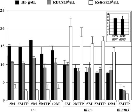
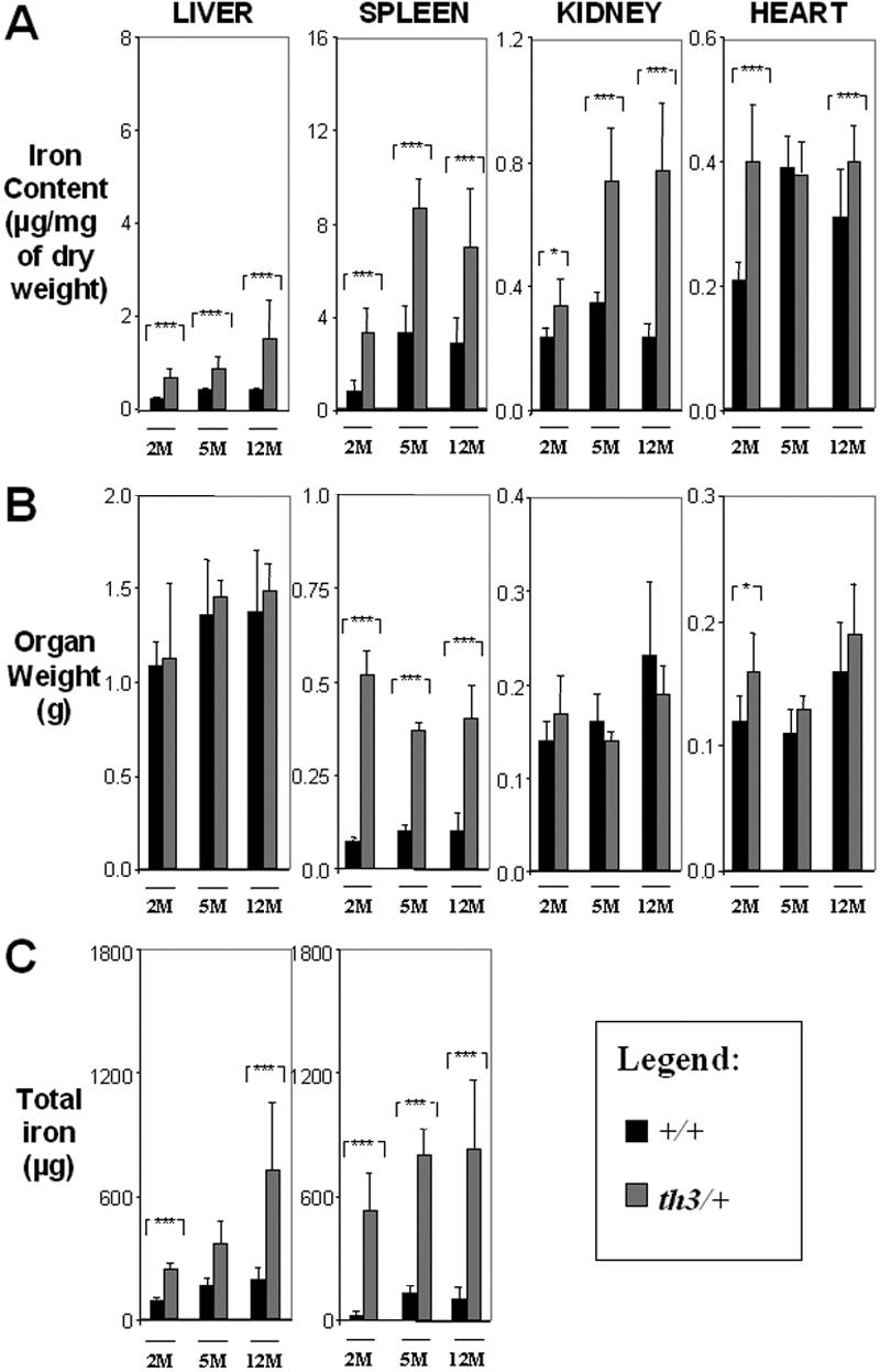
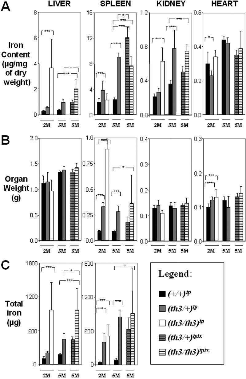
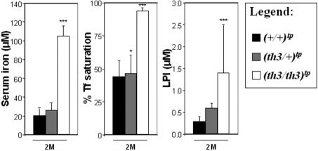
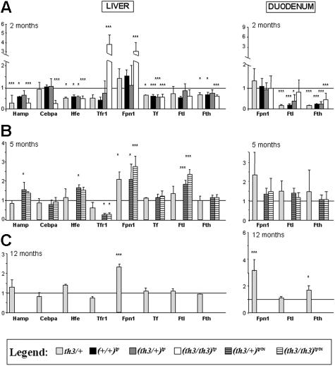
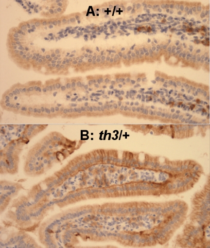
Progressive iron overload is the most salient and ultimately fatal complication of beta-thalassemia. However, little is known about the relationship among ineffective erythropoiesis (IE), the role of iron-regulatory genes, and tissue iron distribution in beta-thalassemia. We analyzed tissue iron content and iron-regulatory gene expression in the liver, duodenum, spleen, bone marrow, kidney, and heart of mice up to 1 year old that exhibit levels of iron overload and anemia consistent with both beta-thalassemia intermedia (th3/+) and major (th3/th3). Here we show, for the first time, that tissue and cellular iron distribution are abnormal and different in th3/+ and th3/th3 mice, and that transfusion therapy can rescue mice affected by beta-thalassemia major and modify both the absorption and distribution of iron. Our study reveals that the degree of IE dictates tissue iron distribution and that IE and iron content regulate hepcidin (Hamp1) and other iron-regulatory genes such as Hfe and Cebpa. In young th3/+ and th3/th3 mice, low Hamp1 levels are responsible for increased iron absorption. However, in 1-year-old th3/+ animals, Hamp1 levels rise and it is rather the increase of ferroportin (Fpn1) that sustains iron accumulation, thus revealing a fundamental role of this iron transporter in the iron overload of beta-thalassemia.


Figures

Figure 1
Figure 1
Mice affected by β-thalassemia intermedia and major show different levels of anemia. The mice have been divided according to their age and treatment (transplantation, tp; blood transfusion, tx). 2M indicates 2-month-old no transplant; 2MTP, 2 months after transplantation; 5M, 5-month-old no transplant; 5MTP, 5 months after transplantation; 12M, 12-month-old no transplant; 5MTPTX, 5 months after transplantation and blood transfusion; ■, Hb (g/dL); ▩, RBC (× 106/μL); □, reticulocytes (× 105/μL). The inset shows Hb levels of (th3/+)tptx-5M and (th3/th3)tptx-5M mice. Error bars represent SD performed on at least 3 animals per group. Sex: female. The age of the animals at transplantation was 8 to 10 weeks.
Figure 2
Figure 2
Organ iron content increases over time in β-thalassemia intermedia. (A) Iron content (μg/mg dry weight), (B) organ weight (g), and (C) total iron (μg) of liver, spleen, kidney, and heart. 2M indicates 2-month-old; 5M, 5-month-old; 12M, 12M-month-old; ■, +/+; ▩, th3/+. Error bars represent SD performed on 3 to 11 independent mice/tissues in duplicate for a total of 90 mice sampled. *P < .05, **P < .01, and ***P < .001 relative to control using the Dunnett multiple comparison test.
Figure 3
Figure 3
Organ iron content differs in β-thalassemia intermedia versus major. (A) Iron content (μg/mg dry weight), (B) organ weight (g), and (C) total iron (μg) of liver, spleen, kidney, and heart. The mice have been categorized according to their age as indicated in Figure 2. Mice receiving transplants are ■, +/+; ▩, th3/+; □, th3/th3; gray striped, th3/+ transfusion; white striped, th3/th3 transfusion. Error bars represent SD performed on 3 to 11 independent mice/tissue in duplicate per group for 90 mice sampled. *P < .05, **P < .01, and ***P < .001 relative to control using the Dunnett multiple comparison test. In the heart, the iron content did not increase in mice given a transplant compared to those not so treated (Figure 2A) and comparing (th3/th3)tp-2M to (th3/+)tp-2M. This indicates that, at least in mice, increasing the body iron content by augmented intestinal absorption or by indirect administration of iron through blood transfusion does not lead to increased accumulation of iron in this organ over time. This was also confirmed by Prussian blue staining (not shown).
Figure 4
Figure 4
Serum iron levels, percentage of Tf saturation, and LPI levels are elevated in mice affected by β-thalassemia major. Mice affected by β-thalassemia major showed a predominant iron deposition in hepatic parenchymal cells. To evaluate whether mice affected by β-thalassemia major had elevated levels of NTBI, we investigated mice at 2 months, given transplants or not so treated. These mice showed the same serum iron, percentage of Tf saturation, and LPI values. Therefore, to simplify our graphs, we combined the animals given transplants and not given transplants into a single group for each genotype. Error bars represent SD performed on at least 5 animals per group. *P < .05, **P < .01, and ***P < .001 relative to controls using the Dunnett multiple comparison test.
Figure 5
Figure 5
In older thalassemic mice, Hamp1 expression increases to the level observed in normal mice, whereas Fpn1 is up-regulated. (A) Mice at 2 months of age. We used the average gene expression value of +/+-2M animals to normalize the values of th3/+-2M mice and of (+/+)tp-2M, (th3/+)tp-2M, and (th3/th3)tp-2M mice, as shown by the horizontal line in the figure. We decided to use this value because our iron data clearly indicated that (+/+)tp-2M mice show increased iron content in their spleen compared to +/+-2M animals. Bars represent the average fold change in mRNA expression compared to +/+-2M mice. (B) Mice at 5 months of age and given transfusions. +/+-5M and (+/+)tp-5M mice showed the same iron values (Figures 2–3) indicating that at this point the effects of the transplantation on erythropoiesis and iron metabolism had disappeared. The same was observed comparing th3/+-5M and (th3/+)tp-5M mice. Therefore, to simplify our graphs, we combined the mice given transplants and not so treated into a single group for each genotype (the light gray bars designating, only in this case, both groups receiving transplants and those that did not). Bars represent the average fold change in mRNA expression when compared with the group of (+/+)tp-5M plus +/+-5M mice. Blood transfusion did not appear to have any effect on the expression level of the genes analyzed in the duodenum, but it increased Hamp1 and normalized and normalized Cebpa liver expression levels in (th3/+)tptx-5M and (th3/th3)tptx-5M mice. Accordingly, in the liver of (th3/+)tptx-5M and (th3/th3)tptx-5M mice, Tfr1 decreased compared to the (th3/+)tp-5M - th3/+-5M mice pool and (th3/th3)tp-2M animals not given transfusions. Ftl1 and Fth1 expression levels were also assessed in the heart, but no differences were observed between thalassemic animals receiving transfusions, those not receiving transfusions, and control animals at 2, 5, and 12 months (not shown). (C) Mice at 12 months. Bars represent the average fold change in mRNA expression when compared with control +/+-12M mice. All the expression levels were normalized using oligonucleotides for mouse Gapdh or β-actin RNA. Ftl1 indicates ferritin-light chain; Fth1, ferritin-heavy chain, Tf, transferrin. Hamp1, Hjv, Hfe, Tfr1, Tfr2, and Tf were extremely low or undetectable in duodenum. For the 60 mice sampled, the Q-PCRs were performed on 3 to 7 independent mice/tissues in duplicate for each gene. Error bars represent SD performed on at least 3 animals per group. *P < .05, **P < .01, and ***P < .001 relative to controls using the Dunnett multiple comparison test. The complete list of genes analyzed is indicated in Table 1. Only genes whose expression was statistically different between control and thalassemic organs are described in this figure.
Figure 6
Figure 6
Fpn1 is increased in the duodenum of 12-month-old mice affected by β-thalassemia intermedia. Duodenum of (A) +/+-12M and (B) th3/+-12M mice. This assay confirmed our Q-PCR data that indicated up-regulation of Fpn1 in 1-year-old th3/+ mice compared to +/+ animals. (immunoperoxidase; original magnification, ×66). Images were captured on a Nikon Eclipse E800 microscope (Nikon, Melville, NY) with a Retiga Exi camera (Qimaging, Burnaby, BC, Canada) and a Plan Fluor 20x/0.75 NA objective, then acquired using IP Lab 3.65a software (Scanalytics, Fairfax, VA). Brightness/contrast and color balance were adjusted using Adobe Photoshop 7.0.1 (Adobe Systems, San Jose, CA).

References

    1. Cooley TB, Lee P. A series of cases of splenomegaly in children with anemia and peculiar bone changes. Trans Am Pediat. Soc. 1925;37:29–33.
    1. Giardina PJ, Grady RW. Chelation therapy in beta-thalassemia: an optimistic update. Semin Hematol. 2001;38:360–366. - PubMed
    1. Bannerman RM, Keusch G, Kreimer-Birnbaum M, Vance VK, Vaughan S. Thalassemia intermedia, with iron overload, cardiac failure, diabetes mellitus, hypopituitarism and porphyrinuria. Am J Med. 1967;42:476–486. - PubMed
    1. Heinrich HC, Gabbe EE, Oppitz KH, et al. Absorption of inorganic and food iron in children with heterozygous and homozygous beta-thalassemia. Z Kinderheilkd. 1973;115:1–22. - PubMed
    1. Cossu P, Toccafondi C, Vardeu F, et al. Iron overload and desferrioxamine chelation therapy in beta-thalassemia intermedia. Eur J Pediatr. 1981;137:267–271. - PubMed

Publication types