Comparative Study
. 2007 Feb 14;2(2):e201.
doi: 10.1371/journal.pone.0000201.

Non-metabolic membrane tubulation and permeability induced by bioactive peptides

Affiliations
Comparative Study

Non-metabolic membrane tubulation and permeability induced by bioactive peptides

Antonin Lamazière et al. PLoS One. .

Abstract

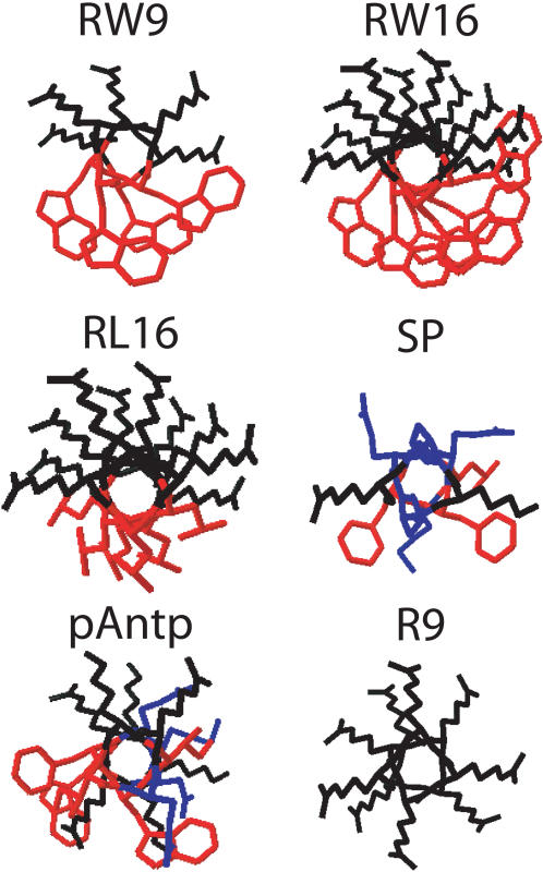
Background: Basic cell-penetrating peptides are potential vectors for therapeutic molecules and display antimicrobial activity. The peptide-membrane contact is the first step of the sequential processes leading to peptide internalization and cell activity. However, the molecular mechanisms involved in peptide-membrane interaction are not well understood and are frequently controversial. Herein, we compared the membrane activities of six basic peptides with different size, charge density and amphipaticity: Two cell-penetrating peptides (penetratin and R9), three amphipathic peptides and the neuromodulator substance P.

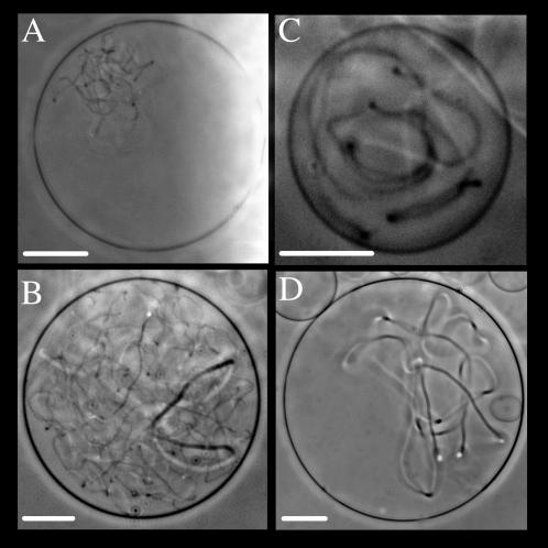
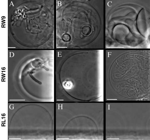
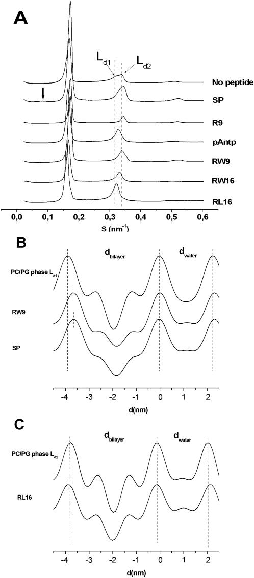
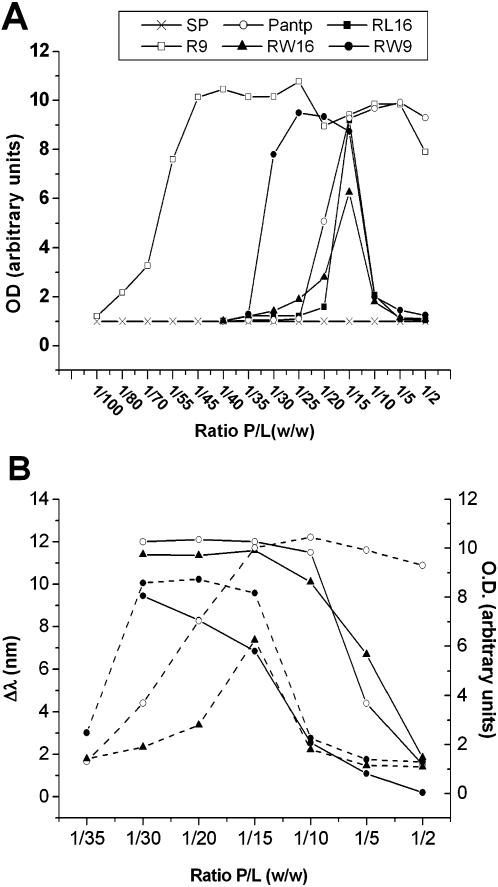
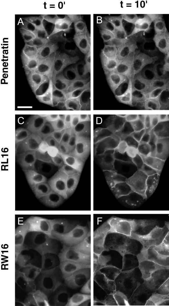
Methodology/principal findings: Experiments of X ray diffraction, video-microscopy of giant vesicles, fluorescence spectroscopy, turbidimetry and calcein leakage from large vesicles are reported. Permeability and toxicity experiments were performed on cultured cells. The peptides showed differences in bilayer thickness perturbations, vesicles aggregation and local bending properties which form lipidic tubular structures. These structures invade the vesicle lumen in the absence of exogenous energy.

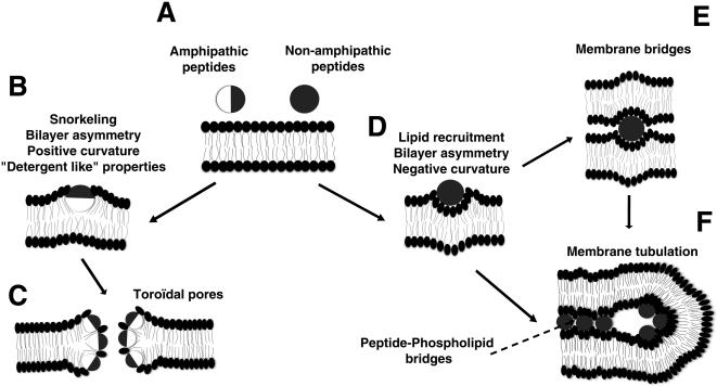
Conclusions/significance: We showed that the degree of membrane permeabilization with amphipathic peptides is dependent on both peptide size and hydrophobic nature of the residues. We propose a model for peptide-induced membrane perturbations that explains the differences in peptide membrane activities and suggests the existence of a facilitated "physical endocytosis," which represents a new pathway for peptide cellular internalization.


Conflict of interest statement

Competing Interests: The authors have declared that no competing interests exist.

Figures

Figure 1
Figure 1
Projections of α–helices of the six basic studied peptides. Basic residues in black, hydrophobic residues in red, other residues in blue. The structure of SP associated to membranes is not known. pAntp is only ∼70% helical when interacting with membranes. The basic/hydrophobic surfaces ratio of RL16 is higher than that of RW16. Helices were generated with the Swiss-PdbViewer programme.
Figure 2
Figure 2
Membrane deformations on GUVs (PC/PG 9/1) induced by two CPPs. Tubular structures observed in vesicles incubated with R9 peptide (A, B), and pAntp (C, D). Phase contrast microscopy at 25°C. Scale bar 20 µm.
Figure 3
Figure 3
Membrane deformations on GUVs (PC/PG 9/1) induced by amphipathic peptides. Coexistence of tubes and small vesicles inside the GUVs (A, B) and adhesion of GUVs by RW9 (C). Tubes formation (D) membrane aggregates (E) and GUVs adhesion and internal vesicles (F) induced by RW16 peptide. Time-lapse sequence of a GUV burst induced by RL16, t = 0 s (G) t = 2 s (H) and t = 3 s (I). Scale bar 10 µm.
Figure 4
Figure 4
Membrane thickness alteration induced by peptides. A) Diffractograms of PC/PG (9/1) MLVs in the absence or presence of peptides with a weight ratio peptide/lipid of 1/20 (at 20°C). Thick arrow shows the position of a weak non lamellar contribution induced by substance P. Ld1 and Ld2 are two lamellar phases clearly distinguished by slightly different d-spacings. B) Electron density profiles of Ld1 phase of peptide free MLVs or formed in the presence of RW9 and SP peptides. C) Electron density profiles of Ld2 phase of peptide free MLVs or formed in the presence of RW16 and RL16.
Figure 5
Figure 5
Membrane bridging capacity of peptides. A) Aggregation profiles of PC/PG (9/1) vesicles at different Peptide/Lipids weight ratios: R9 (○), pAntp (□), RW9 (•), RW16 (▪), RL16 (▴) and SP (X). Optical density (OD) was recorded at the plateau of aggregation (20 min after peptide addition). B) Membrane binding and aggregation of RW16, RW9 and pAntp peptides. Shift in emission wavelength (λ shift) in function of P/L ratio (solid lines). LUVs aggregation (dotted lines). For presentation facility, data were adjusted in arbitrary units. pAntp (□), RW9 (•), RW16 (▪). All experiments were achieved at 25°C.
Figure 6
Figure 6
Peptide-induced LUVs permeability at different Peptide/Lipid weight ratio. Percent of calcein release from LUVs 5 min after peptide addition: R9 (○), pAntp (□), RW9 (•), RW16 (▪), RL16 (▴) and SP (X). Experiments were achieved at 25°C.
Figure 7
Figure 7
Movement of Annexin 2-GFP by peptide-induced ion permeabilization of the plasma membrane. Visualisation by fluorescence microscopy of the Anx2-GFP migration from the cytsol to the plasma membrane before and after addition of peptides (10 µM). pAntp, RL16 and RW16. (A,C,E before peptide addition, B,D,F 10 min after peptide addition). Scale bar 10 µm.
Figure 8
Figure 8
Model for peptide-membrane interaction. A) Two main properties are considered: amphipathicity and net positive charge. Changes in these properties result in different degrees of membrane perturbations. Basic surface in dark, hydrophobic surface in white. B) The binding of the amphipathic peptides involves electrostatic interactions between the basic residues and lipid headgroup negative charges, and hydrophobic interactions with lipid fatty acyl moieties. A strong snorkelling effect of the peptide could induce protrusion of headgroups attracted by the charged helix residues inducing lipid reorganisation resulting in asymmetry of the bilayer halves and positive curvature of the membrane. C) The detergent property of amphipathic peptides and the induced positive curvature result in the formation of toroidal pores. D) The binding of basic peptides mainly involves electrostatic interactions with the lipid headgroup phosphates. This results in the recruitment of phospholipids and then in membrane asymmetry with negative curvature. E) The bridging interaction of the peptide between two membranes allows adhesion and vesicles aggregation. F) Membrane tubulation results from the membrane curvature inducing invaginations. The thin tubes could be stabilized by the bridging properties of the peptides between membranes apposed faces.

References

    1. Dietz GP, Bahr M. Delivery of bioactive molecules into the cell: the Trojan horse approach. Mol Cell Neurosci. 2004;27:85–131. - PubMed
    1. Nekhotiaeva N, Elmquist A, Rajarao GK, Hallbrink M, Langel U, et al. Cell entry and antimicrobial properties of eukaryotic cell-penetrating peptides. Faseb J. 2004;18:394–396. - PubMed
    1. Palm C, Netzereab S, Hallbrink M. Quantitatively determined uptake of cell-penetrating peptides in non-mammalian cells with an evaluation of degradation and antimicrobial effects. Peptides. 2006;27:1710–1716. - PubMed
    1. Duplaa H, Convert O, Sautereau AM, Tocanne JF, Chassaing G. Binding of substance P to monolayers and vesicles made of phosphatidylcholine and/or phosphatidylserine. Biochim Biophys Acta. 1992;1107:12–22. - PubMed
    1. Oehlke J, Lorenz D, Wiesner B, Bienert M. Studies on the cellular uptake of substance P and lysine-rich, KLA-derived model peptides. J Mol Recognit. 2005;18:50–59. - PubMed

Publication types

MeSH terms