. 2007 Jun 1;404(2):235-45.
doi: 10.1042/BJ20061830.

Mice develop normally in the absence of Smad4 nucleocytoplasmic shuttling

Affiliations

Mice develop normally in the absence of Smad4 nucleocytoplasmic shuttling

Christine A Biondi et al. Biochem J. .

Abstract

Smad4 in partnership with R-Smads (receptor-regulated Smads) activates TGF-beta (transforming growth factor-beta)-dependent signalling pathways essential for early mouse development. Smad4 null embryos die shortly after implantation due to severe defects in cell proliferation and visceral endoderm differentiation. In the basal state, Smad4 undergoes continuous shuttling between the cytoplasm and the nucleus due to the combined activities of an N-terminal NLS (nuclear localization signal) and an NES (nuclear export signal) located in its linker region. Cell culture experiments suggest that Smad4 nucleocytoplasmic shuttling plays an important role in TGF-beta signalling. In the present study we have investigated the role of Smad4 shuttling in vivo using gene targeting to engineer two independent mutations designed to eliminate Smad4 nuclear export. As predicted this results in increased levels of Smad4 in the nucleus of homozygous ES cells (embryonic stem cells) and primary keratinocytes, in the presence or absence of ligand. Neither mutation affects Smad4 expression levels nor its ability to mediate transcriptional activation in homozygous cell lines. Remarkably mouse mutants lacking the Smad4 NES develop normally. Smad4 NES mutants carrying one copy of a Smad4 null allele also fail to display developmental defects. The present study clearly demonstrates that Smad4 nucleocytoplasmic shuttling is not required for embryonic development or tissue homoeostasis in normal, healthy adult mice.


Figures
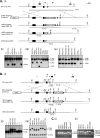
Figure 1
Figure 1. Generation of Smad4ΔNES and Smad4MEX alleles
Gene targeting strategies to (A) flank exon 4 containing the NES with loxP sites (large arrows) or (B) insert a mutated exon 4 into the endogenous Smad4 locus are shown. For both constructs, hygromycin-resistant targeted clones were screened with a 3′ external probe (probe a; grey box) to identify homologous recombinants. (A) ii, Following electroporation, targeted clones which had incorporated the targeting construct including the intron 4 loxP site (Smad4ΔNES−TA/+) were distinguished from random integration event clones (Smad4+/+) by screening DNA digested with BstZ17I with probe a. The positions of the 9.4 kb WT (wild-type) and 5.2 kb TA (targeted allele) bands are indicated. iii, For excision of the selection cassette, Smad4ΔNES−TA/+ clones were subjected to transient transfection with Cre recombinase. Screening with probe b [shown as a black box on (A, i)], confirmed excision of the hygro cassette and exon 4 (Smad4ΔNES/+; 8.6 kb), the hygro cassette only (i.e. conditional allele Smad4ΔNES−CA/+; 4.2 kb) or retention of the Smad4ΔNES−TA/+ allele (6.4 kb). iv, Smad4ΔNES/+ sub-clones were retargeted and screened with probe a, to detect either the Smad4ΔNES−TA (5.2 kb), Smad4ΔNES (8.6 kb) or Smad4+/+ (9.4 kb) alleles. Clones in which the remaining Smad4 wild-type allele had been disrupted (i.e. Smad4ΔNES−TA/ΔNES; 5.2 and 8.6 kb) were then subjected to in vitro Cre-mediated excision to generate homozygous mutant cell lines lacking both the pgk-hygro cassette and exon 4 (results not shown). (B) ii, Following electroporation, correctly targeted Smad4MEX−TA/+ clones were identified by screening BstZ17I-digested DNA with probe a. The position of the 11.6 kb TA band is indicated. Further analysis via PstI digestion and screening with probe c was used to identify clones that had incorporated the mutant exon 4 and hence lost the endogenous PstI site (Smad4MEX−TA/+,1.3 kb or Smad4+/+, 0.75 kb). iii, Screening of PstI digested DNA from Smad4MEX−TA/+ ES clones following transient transfection with pMC1-Cre. Probe c [shown as a black box on (B, i)] distinguishes Smad4MEX/+ (1.4 kb), Smad4MEX−TA/+ (1.3 kb) or Smad4+/+ (0.75 kb) clones. Bz, BstZ17I; Bs, BspE1 and Ps, PstI. (C) PCR genotyping screen. i, Amplification of genomic DNA between primers Ex2F and Int3R (small arrows) detects three possible alleles, Smad4+, Smad4ΔNES and Smad4ΔNES−CA (1.0, 0.4 and 1.1 kb respectively). ii, To detect the Smad4MEX allele, PCR products generated using the same primers were digested with PstI to generate diagnostic banding patterns for wild-type (560, 270 and 175 bp), Smad4MEX/+ (830, 560, 270 and 175 bp) and Smad4MEX/MEX (830 and 175 bp).
Figure 2
Figure 2. Analysis of mRNA and protein expression in homozygous mutant ES cells
(AC) RPA studies. (A) Total RNA was hybridized with probes specific for representative regions of the Smad4 cDNA as illustrated. (B) Probe A (exons 4–5) distinguishes Smad4 full-length and the ΔNES isoform. Both alternative splice variants are co-expressed in Smad4+/+ ES cells. Expression of ΔNES transcripts is up-regulated in heterozygous ES cells (Smad4ΔNES/+; 3C11 and 3B8) and the 243 full-length product is absent in homozygous mutant ES cells (Smad4ΔNES/ΔNES, NES#1 and #5). The sizes of the protected fragments and expression ratios are indicated. (C) Analysis of downstream splicing events. Protected fragments detectable with probes B (exons 5–9) and C (exons 9–10) are present at the same levels irrespective of the genotype (lower panels). Thus the deletion has no detectable effect on splicing ratios within the linker or further downstream within the MH2 domain. (D) Homozygous mutant ES cells produce Smad4 protein lacking exon 4. Western analysis reveals that Smad4ΔNES/ΔNES ES cells (NES#1 and #5) generate Smad4 protein at wild-type levels. In contrast to full-length protein detected in Smad4+/+ ES cell lines (CCE; upper panel), Smad4N/N cells (BNN), lack Smad4 protein, whereas the Smad4ΔNES (NES#1 and #5) product shows a slightly faster mobility. Expression of R-Smads (i.e. Smad1, 2 and 3) is unaffected. Blots were probed with anti-Smad4 and anti-Smad2/3 mouse monoclonal antibodies and anti-Smad1 and anti-Smad3 rabbit polyclonal antibodies. Grb2 is included as a loading control (bottom panel).
Figure 3
Figure 3. Nuclear localization of Smad4 in ES cells lacking the NES
Nucleocytoplasmic distribution of Smad proteins in ES cells. ES cells were grown on gelatin-coated coverslips under normal conditions (uninduced); treated with SB-431542 overnight followed by TGF-β induction for 1 h prior to fixation (+TGF-β). Cells were fixed and then processed for immunofluorescence using mouse monoclonal antibodies against either (A) Smad4 or (B) Smad2/3. (A) Smad4 shifts from a predominantly cytoplasmic localization in wild-type cells (i) to a largely nuclear localization in Smad4ΔNES/ΔNES mutants (ii and iii). Following ligand stimulation, wild-type Smad4 rapidly accumulates in the nucleus and exhibits a predominantly nuclear staining pattern (iv), similar to that observed in the ΔNES mutants (v and vi). (B) Smad2/3 is broadly distributed in cells expressing all three genotypes. Responsiveness to ligand stimulation results in nuclear Smad2/3 accumulation (iv–vi). Indistinguishable localization patterns were observed for Smad4ΔNES/ΔNES ES cell lines, NES #7 and #8 in independent experiments. Fluorescence was visualised using a Zeiss LSM 510 confocal microscope. Representative images are shown in green (upper panels) with DAPI nuclear counter-staining (blue) of the same cells provided in the lower panels. CCE, Smad4+/+ (i and iv); NES#1, Smad4ΔNES/ΔNES (ii and v) and NES#5, Smad4ΔNES/ΔNES (iii and vi).
Figure 4
Figure 4. Targeted disruption of the Smad4 nuclear localization signal restricts nucleocytoplasmic distribution in keratinocytes
Keratinocytes were grown on collagen-coated coverslips for 24 h in growth factor starvation medium (uninduced). Some cells were then treated for 1 h with TGF-β or LMB, as indicated, prior to fixation. Cells were then stained with either (A) Smad4 or (B) Smad2/3 monoclonal antibodies. (A) Smad4 shifts from a predominantly cytoplasmic localization in wild-type cells (i) to a largely nuclear localization in Smad4ΔNES/ΔNES and Smad4MEX/MEX mutants (ii and iii). Following ligand stimulation, wild-type Smad4 rapidly accumulates in the nucleus (iv), similar to that observed in the Smad4ΔNES/ΔNES and Smad4MEX/MEX mutants (v and vi). LMB treatment of wild-type cells results in accumulation of Smad4 in the nucleus (vii) in a similar pattern to cells lacking a functional NES i.e. Smad4ΔNES/ΔNES and Smad4MEX/MEX mutants (viii and ix). (B) For all three genotypes Smad2/3 was distributed throughout the cell (i–iii) and stimulation with TGF-β results in Smad2/3 nuclear localization. (iv–vi). LMB treatment has no effect on Smad2/3 nucleocytoplasmic distribution (vii–ix). Fluorescence was visualised using a Zeiss LSM 510 confocal microscope. Representative images are shown in green (upper panels) and DAPI nuclear counter-staining (blue) of the same cells is provided in the lower panels.
Figure 5
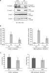
Figure 5. Smad4ΔNES efficiently forms transcriptionally active complexes with phosphorylated Smad2 and Smad3
(A) Smad4ΔNES co-precipitates with phosphorylated-Smad2 (P-Smad2). Smad4 complexes were immunoprecipitated and Western blots subsequently probed with an anti-phosphorylated-Smad2 antibody. Control cell extracts from Smad4ΔNES/ΔNES or Smad4+/+ MEFs were Western blotted for phosphorylated-Smad2, total Smad2 or Smad4 as indicated. Cells were cultured with 10 μM SB-431542 (SB) overnight to abolish autocrine signalling, and then washed twice with PBS and treated with 2 ng/ml of TGF-β (+TGF-β) for 1 h. (B and C) Smad4ΔNES efficiently functions as a transcriptional activator. Smad4ΔNES/ΔNES or Smad4+/+ MEFs were transfected with CAGA12-luciferase and EF-LacZ as a control, or with ARE3-luciferase, EF-Flag-XFoxH1a and EF-LacZ. Cells pre-treated with SB-431542 overnight, as in (A), were washed and induced with TGF-β for 8 h or kept in SB-431542. Luciferase activity was quantified relative to β-galactosidase activity. The normalized luciferase values±TGF-β are shown in (B) and the fold inductions are shown in (C). The data are the average of triplicates. The values for the Smad4ΔNES/ΔNES MEFs are all lower than those for the Smad4+/+ MEFs. This is a result of the transfection efficiency being lower for the Smad4ΔNES/ΔNES MEFs compared to the Smad4+/+ MEFs. However, the fold inductions are the same.

Similar articles

Cited by

References

    1. Shi Y., Massague J. Mechanisms of TGF-β signaling from cell membrane to the nucleus. Cell. 2003;113:685–700. - PubMed
    1. Massague J., Seoane J., Wotton D. Smad transcription factors. Genes Dev. 2005;19:2783–2810. - PubMed
    1. Liu F., Pouponnot C., Massagué J. Dual role of the Smad4/DPC4 tumor suppressor in TGFβ-inducible transcriptional complexes. Genes Dev. 1997;11:3157–3167. - PMC - PubMed
    1. Reguly T., Wrana J. L. In or out? The dynamics of Smad nucleocytoplasmic shuttling. Trends Cell Biol. 2003;13:216–220. - PubMed
    1. Xu L., Kang Y., Col S., Massague J. Smad2 nucleocytoplasmic shuttling by nucleoporins CAN/Nup214 and Nup153 feeds TGFβ signaling complexes in the cytoplasm and nucleus. Mol. Cell. 2002;10:271–282. - PubMed

Publication types