. 2007 Mar;17(3):209-20.
doi: 10.1016/j.nmd.2006.12.004. Epub 2007 Feb 14.

Postnatal overexpression of the CT GalNAc transferase inhibits muscular dystrophy in mdx mice without altering muscle growth or neuromuscular development: evidence for a utrophin-independent mechanism

Affiliations

Postnatal overexpression of the CT GalNAc transferase inhibits muscular dystrophy in mdx mice without altering muscle growth or neuromuscular development: evidence for a utrophin-independent mechanism

Rui Xu et al. Neuromuscul Disord. 2007 Mar.

Abstract

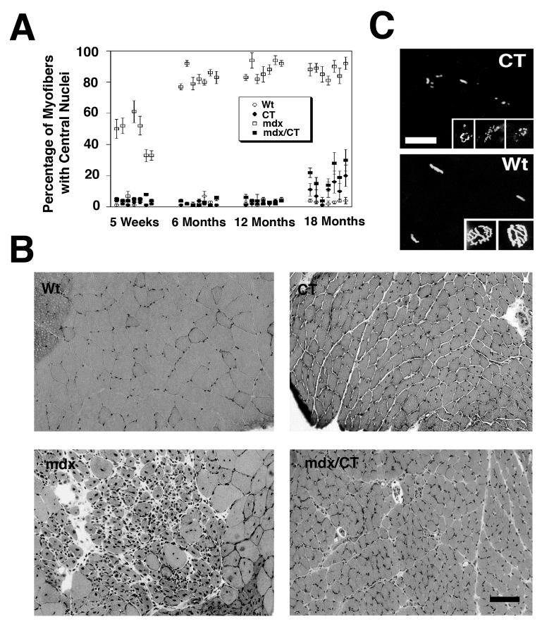
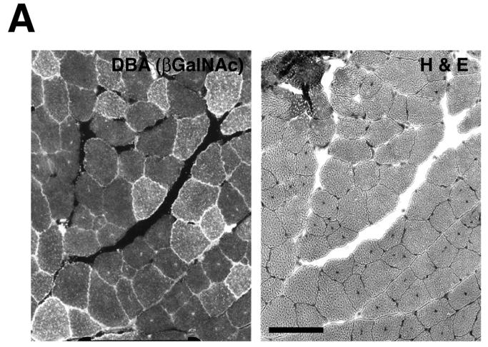
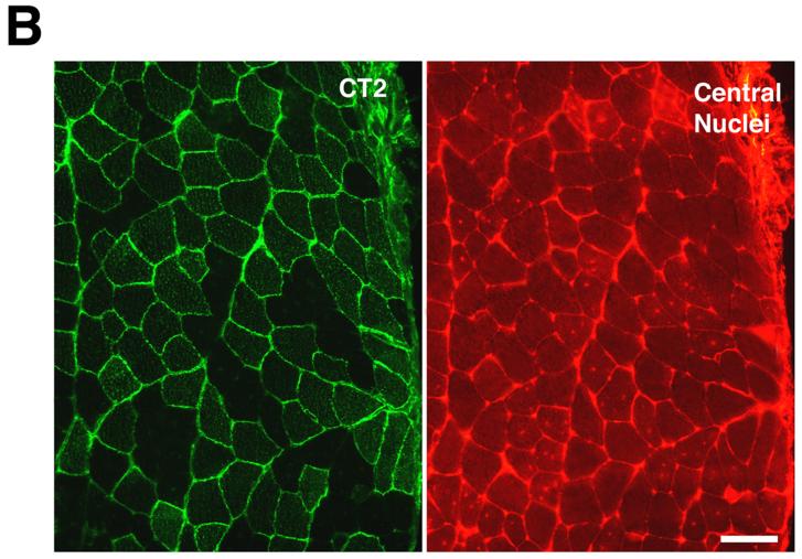
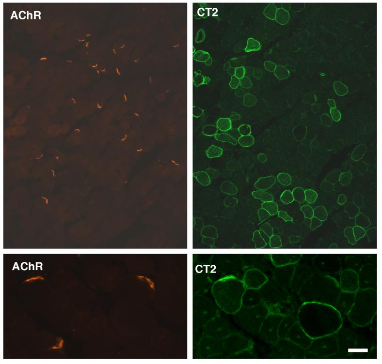
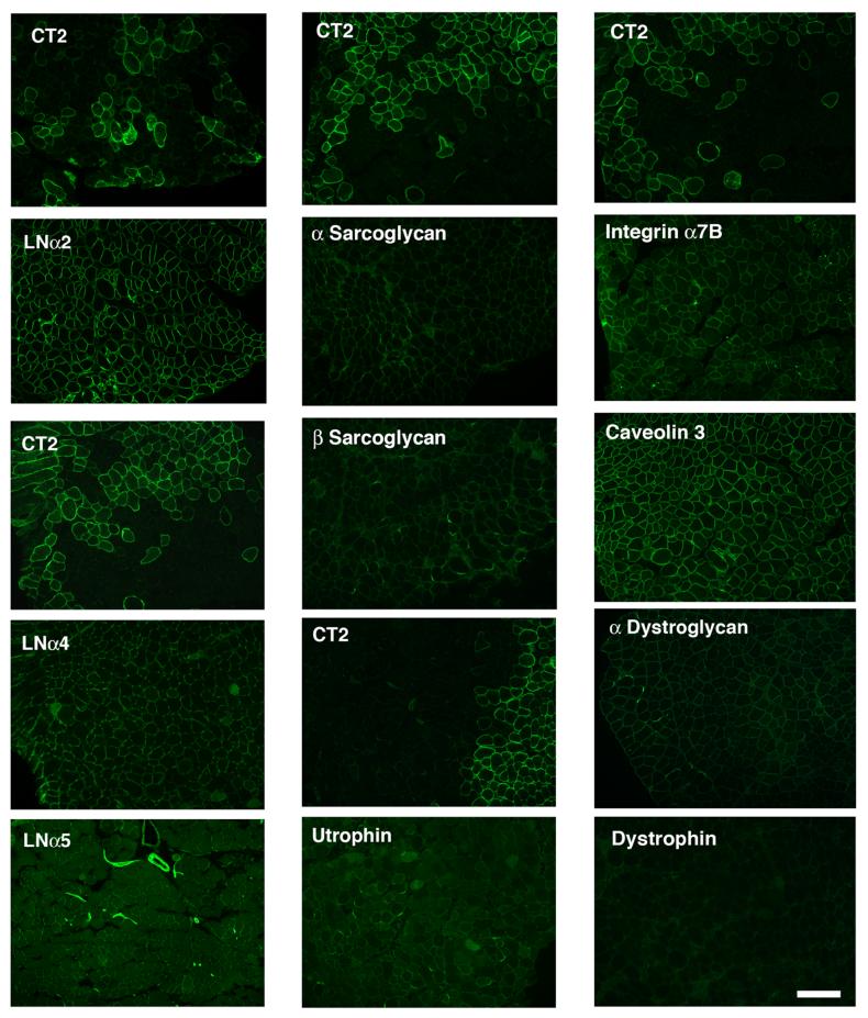
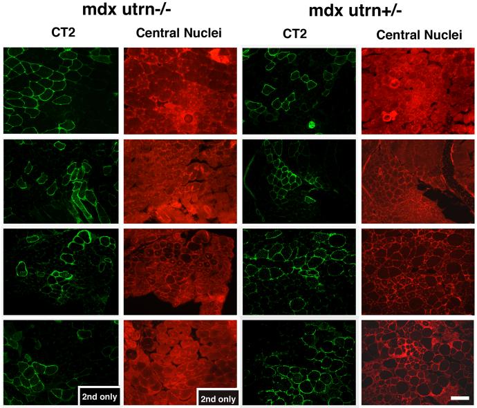
Overexpression of the cytotoxic T cell (CT) GalNAc transferase (Galgt2) in the skeletal muscles of transgenic mdx mice has been reported to inhibit the development of muscular dystrophy. The profound effect of Galgt2 on muscular dystrophy in transgenic mice, where overexpression is begins from embryonic stages, is complicated by its additional effects on muscle growth and neuromuscular structure. Here, we use adeno-associated virus (AAV) to show that overexpression of Galgt2 in skeletal myofibers in the early postnatal period is equally effective in inhibiting muscular dystrophy, but that it does so without altering muscle growth or neuromuscular structure. Unlike embryonic overexpression, postnatal overexpression of Galgt2 did not reproducibly increase the expression of utrophin, synaptic laminins, or dystrophin-associated glycoproteins along infected myofibers. Moreover, Galgt2 overexpression inhibited muscular dystrophy to the same extent in utrophin-deficient mdx muscles as it did in utrophin-expressing mdx muscles. Thus, Galgt2 is a molecular target for therapy in DMD that can be utilized in a manner that separates its clinical benefit from its effects on development, and its clinical benefit is distinct from that achieved by utrophin.


Figures

Figure 1
Figure 1
Overexpression of CT carbohydrate inhibits muscle pathology up to 18 months of age in Galgt2 transgenic mdx mice. (A) Central nuclei were quantitated in skeletal muscles taken from wild type (Wt), Galgt2 transgenic (CT), mdx, and Galgt2-transgenic mdx (mdx/CT) mice at the indicated ages. Skeletal muscles analyzed (from left to right) were gastrocnemius, trapezius, diaphragm, triceps, quadriceps, tibialis anterior, and gluteus maximus. CT muscles began to develop some central nuclei at 18 months of age, as did mdx/CT muscles, however, the percentage of myofibers with central nuclei in mdx/CT animals was still significantly lower than mdx littermates for all muscles (P<0.001 for all) at 12 and 18 months of age. Errors are SEM for n=250 fibers per muscle for 3-4 animals per condition. (B) Hematoxylin and eosin staining of muscles at 18 months of age showed that CT and mdx/CT myofibers were significantly smaller than wild type (gastrocnemius is shown) and relatively free from muscle damage. Bar is 50μm. (C) Rhodamine-α-bungarotoxin of Wt and CT muscles in cross-section (larger panels) or in whole mount (smaller panels) shows aberrant neuromuscular junctions in CT muscles as compared to wild type at 18 months of age as well. Bar is 25μm (larger panels) or 12.5μm (smaller panels).
Figure 2
Figure 2
Postnatal overexpression of CT carbohydrate in mdx skeletal muscle inhibits the development of muscle pathology but not muscle growth. (A) AAV2-Galgt2 was used to overexpress CT carbohydrate in mdx muscle (tibialis anterior, infection at 2 weeks, staining at 6 weeks). Muscle was stained with DBA to identify CT carbohydrate overexpressing myofibers (left) and with hematoxylin and eosin on the next section in a serial series to determine myofibers with central nuclei. Myofibers overexpressing CT carbohydrate were protected from developing central nuclei. (B) Gastrocnemius muscle in an mdx mouse was infected with AAV1-Galgt2 at 2 weeks of age and analyzed for CT carbohydrate overexpression (green) and for central nuclei (red) at 6 weeks of age. Myofibers overexpressing CT carbohydrate were protected from muscle damage and did not contain central nuclei. Bar is 50μm in A and B.
Figure 2
Figure 2
Postnatal overexpression of CT carbohydrate in mdx skeletal muscle inhibits the development of muscle pathology but not muscle growth. (A) AAV2-Galgt2 was used to overexpress CT carbohydrate in mdx muscle (tibialis anterior, infection at 2 weeks, staining at 6 weeks). Muscle was stained with DBA to identify CT carbohydrate overexpressing myofibers (left) and with hematoxylin and eosin on the next section in a serial series to determine myofibers with central nuclei. Myofibers overexpressing CT carbohydrate were protected from developing central nuclei. (B) Gastrocnemius muscle in an mdx mouse was infected with AAV1-Galgt2 at 2 weeks of age and analyzed for CT carbohydrate overexpression (green) and for central nuclei (red) at 6 weeks of age. Myofibers overexpressing CT carbohydrate were protected from muscle damage and did not contain central nuclei. Bar is 50μm in A and B.
Figure 3
Figure 3
Neuromuscular junctions in mdx myofibers appear normal after postnatal overexpression of CT carbohydrate. Mdx muscles were infected with AAV1-Galgt2 at 2 weeks of age and analyzed at 6 weeks. Muscles were co-stained for CT carbohydrate overexpression (using the CT2 monoclonal antibody, right) and with rhodamine-α-bungarotoxin (left) to label nicotinic acetylcholine receptors at the neuromuscular junction. Neuromuscular junctions appear normal in mdx muscles where CT carbohydrate is overexpressed. Bar is 40μm (top panels) and 10μm (bottom panels).
Figure 4
Figure 4
Expression of utrophin, synaptic laminins, and dystrophin-associated glycoproteins is not increased with postnatal overexpression of the CT carbohydrate in mdx skeletal muscles. Mdx muscles were infected with AAV1-Galgt2 at 1-2 weeks of age and analyzed at 6 weeks. Serial sections were stained with antibodies to the CT carbohydrate (CT2) or with antibodies to the indicated proteins (panels below CT2 panels). Overexpression of the CT carbohydrate in postnatal mdx skeletal muscle did not increase expression of utrophin, dystroglycan, sarcoglycans, laminin α4, laminin α5, or integrin α7B along CT-overexpressing myofibers. Bar is 50μm.
Figure 5
Figure 5
Postnatal overexpression of Galgt2 does not increase CT glycosylation of α dystroglycan or expression of utrophin, synaptic laminins, or dystrophin-associated glycoproteins in mdx muscle. Left, 150μg of NP-40 detergent protein lysate was precipitated with a GalNAc-binding lectin that can identify the CT carbohydrate (WFA agarose) or with a control lectin known to precipitate α dystroglycan (WGA agarose). Proteins were eluted with GalNAc (for WFA) or GlcNAc (for WGA) and analyzed by Western blot for α dystroglycan or CT carbohydrate (CT2). Postnatal overexpression of Galgt2 using AAV caused a slight increase in α dystroglycan precipitated with WFA, but this protein was not glycosylated with the CT carbohydrate. By contrast, WFA precipitated CT-glycosylated α dystroglycan from Galgt2 transgenic mdx muscle (mdx/CT). WGA precipitation showed equal amounts of α dystroglycan in each pair of samples. Right, comparison of whole cell muscle lysates showed postnatal overexpression of Galgt2 did not increase expression of utrophin, α dystroglycan, β dystroglycan, laminin α2, laminin α4, laminin α5, α sarcoglycan, or β sarcoglycan protein. Blots were stripped and reprobed for actin as a control for protein loading and transfer.
Figure 6
Figure 6
Postnatal overexpression of Galgt2 inhibits muscular dystrophy in mdx mice lacking utrophin. Mdx mice lacking utrophin (mdxutrn-/-) or expressing utrophin (mdxutrn+/-) were infected with AAV1-Galgt2 or AAV8-like (rh.74) Galgt2 at 4 days to 1 week of age and analyzed at 6 weeks of age. Infected muscles were stained with antibody to the CT carbohydrate (CT2) in the fluorescein channel (green) and with a non-specific antibody to mark central nuclei in the rhodamine channel (red). Mdx myofibers overexpressing the CT carbohydrate did not have central nuclei, regardless of utrophin expression. Bar is 50μm.

References

    1. Hoffman EP, Brown RH, Jr., Kunkel LM. Dystrophin: the protein product of the Duchenne muscular dystrophy locus. Cell. 1987;51:919–928. - PubMed
    1. Koenig M, Hoffman EP, Bertelson CJ, Monaco AP, Feener C, Kunkel LM. Complete cloning of the Duchenne muscular dystrophy (DMD) cDNA and preliminary genomic organization of the DMD gene in normal and affected individuals. Cell. 1987;50:509–517. - PubMed
    1. Blake DJ, Weir A, Newey SE, Davies KE. Function and genetics of dystrophin and dystrophin-related proteins in muscle. Physiol Rev. 2002;82:291–329. - PubMed
    1. De la Porte S, Morin S, Koenig J. Characteristics of skeletal muscle in mdx mutant mice. Int Rev Cytol. 1999;191:99–148. - PubMed
    1. Matsumura K, Ervasti JM, Ohlendieck K, Kahl SD, Campbell KP. Association of dystrophin-related protein with dystrophin-associated proteins in mdx mouse muscle. Nature. 1992;360:588–591. - PubMed

Publication types