Comparative Study
. 2007 May;81(9):4473-81.
doi: 10.1128/JVI.02498-06. Epub 2007 Feb 14.

Rotavirus NSP1 inhibits expression of type I interferon by antagonizing the function of interferon regulatory factors IRF3, IRF5, and IRF7

Affiliations
Comparative Study

Rotavirus NSP1 inhibits expression of type I interferon by antagonizing the function of interferon regulatory factors IRF3, IRF5, and IRF7

Mario Barro et al. J Virol. 2007 May.

Abstract

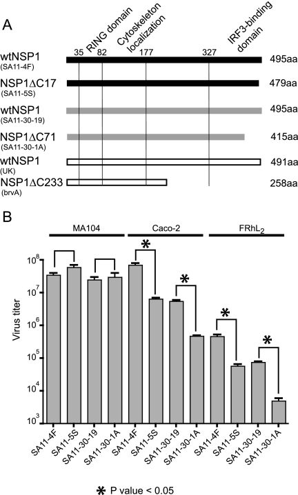
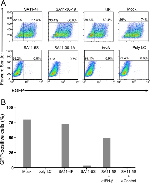
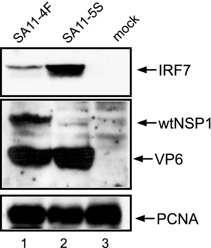
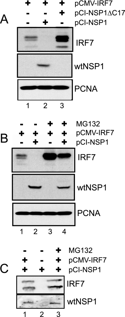
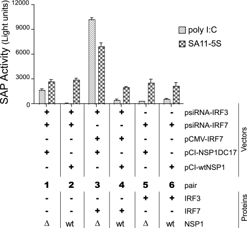
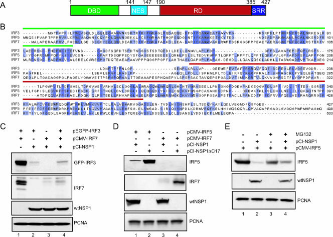
Secretion of interferon (IFN) by virus-infected cells is essential for activating autocrine and paracrine pathways that promote cellular transition to an antiviral state. In most mammalian cells, IFN production is initiated by the activation of constitutively expressed IFN regulatory factor 3, IRF3, which in turn leads to the induction of IRF7, the "master regulator" of IFN type I synthesis (alpha/beta IFN). Previous studies established that rotavirus NSP1 antagonizes IFN signaling by inducing IRF3 degradation. In the present study, we have determined that, in comparison to wild-type rotaviruses, rotaviruses encoding defective NSP1 grow to lower titers in some cell lines and that this poor growth phenotype is due to their failure to suppress IFN expression. Furthermore, we provide evidence that rotaviruses encoding wild-type NSP1 subvert IFN signaling by inducing the degradation of not only IRF3, but also IRF7, with both events occurring through proteasome-dependent processes that proceed with similar efficiencies. The capacity of NSP1 to induce IRF7 degradation may allow rotavirus to move across the gut barrier by enabling the virus to replicate in specialized trafficking cells (dendritic cells and macrophages) that constitutively express IRF7. Along with IRF3 and IRF7, NSP1 was found to induce the degradation of IRF5, a factor that upregulates IFN expression and that is involved in triggering apoptosis during viral infection. Our analysis suggests that NSP1 mediates the degradation of IRF3, IRF5, and IRF7 by recognizing a common element of IRF proteins, thereby allowing NSP1 to act as a broad-spectrum antagonist of IRF function.


Figures

FIG. 1.
FIG. 1.
In vitro growth of rotaviruses encoding wt and defective forms of NSP1. (A) Representation of the NSP1 forms made by different rotavirus isolates (strain name in parentheses), including the location of putative RING and IRF3-binding domains, and cytoskeleton localization signal. The variants SA11-5S, SA11-30-1A, and brvA encode C-truncated forms of NSP1 and are sister strains of the wt viruses SA11-4F (SA11g4“O”), SA11-30-19, and bovine UK (, a). GenBank accession numbers for the gene 5 sequences: AAK14071 (SA11-4F), AAK14072 (SA11-5S), AAK14069 (SA11-30-19), AAK14070 (SA11-30-1A), AAA18011 (UK), and AAA18012 (brvA). (B) Virus titers produced by infecting the indicated cell lines at an MOI of 3 with rotavirus strains encoding wt and defective NSP1. Cells were harvest at times of maximum virus yield (24 h p.i. for MA104 cells and 48 h p.i. for Caco-2 and FRhL2 cells). Titers were determined by plaque assay on MA104 cells and represent the averaged values obtained for replicate parallel infections.
FIG. 2.
FIG. 2.
Induction of IFN associated with rotavirus infection. (A) Culture media from FRhL2 cells that were mock infected or infected with the indicated strains of rotavirus or treated with poly(I:C) were transferred onto fresh FRhL2 monolayers. After 24 h, the monolayers were infected with VSV-GFP, an IFN-sensitive virus expressing GFP. Flow cytometry was used to analyze cells for GFP fluorescence and cell size. Plots of the data indicate the percentage of cells expressing GFP (upper right quadrant) beyond the background level of fluorescence associated with uninfected control cells. (B) Same as in panel A, except that in some cases, neutralizing IFN-β antisera and control antisera were added to the culture media of SA11-5S-infected cells prior to transfer to fresh FRhL2 monolayers. The monolayers were later infected with VSV-GFP, and the percentage of cells expressing GFP was determined by flow cytometry.
FIG. 3.
FIG. 3.
IRF7 levels in rotavirus-infected cells. Lysates prepared at 18 h p.i. from Caco-2 cells infected with SA11-4F or SA11-5S were examined for IRF7, full-length NSP1, VP6, and PCNA by Western blot assay. The SA11 NSP1(C19) antiserum was prepared against a peptide representing the last 19 amino acids of wt NSP1 (15). As a result, the antiserum does not recognize the C-truncated NSP1 (NSP1ΔC17) encoded by SA11-5S.
FIG. 4.
FIG. 4.
Proteasome-dependent effect of NSP1 on IRF7 levels. (A) 293T cells were cotransfected with equal amounts of pCMV-IRF7 and pCI-NSP1ΔC17 or pCI-NSP1. Where necessary, the empty pCI vector was also added to normalize amounts of plasmid DNA contained in transfection mixtures. Lysates prepared from the cells were examined at 48 h posttransfection for IRF7, wt NSP1, and PCNA by Western blot assay. (B) Same as in panel A, except that at 48 h posttransfection the culture media over some monolayers was adjusted to 10 μM MG132, an inhibitor of proteasome function. Lysates were prepared 12 h later and analyzed by Western blot assay. (C) Same as in panel B, except that immunoprecipitates were recovered by using IRF7-antibody linked to agarose beads from cellular lysates prepared at 24 posttransfection. The immunoprecipitates were analyzed for IRF7 and wt NSP1 by Western blot assay.
FIG. 5.
FIG. 5.
Importance of IRF3 and IRF7 expression on rotavirus growth. (A) Vectors producing IRF7- or IRF3-specific siRNAs (psiRNA-IRF7 and -IRF3, respectively) or a control irrelevant siRNA (psiRNA-scramble) were nucleofected into Caco-2 cells. After incubation with 103 U of IFN-β per ml for 10 h, the cells were analyzed for IRF3, IRF7, and PCNA by Western blot assay. (B) Vectors producing IRF7- or IRF3-specific siRNAs or a control irrelevant siRNA were nucleofected into Caco-2 cells. After 24 h, the cells were infected with SA11-4F or SA11-5S at an MOI of 3. The infected Caco-2 cells were harvested at 48 h p.i., a time by which SA11-4F has reached maximum titers in this cell line. Virus titers were determined by plaque assay on MA104 cells. The titers shown reflect the averaged results of two separate experiments, with plaque assays performed in duplicate.
FIG. 6.
FIG. 6.
Effect of NSP1 on the activation of the IFN-α promoter element. Caco-2 cells were nucleofected with combinations of vectors expressing IRF3- or IRF7-specific siRNAs, wt NSP1 or NSP1ΔC17, IRF7, and pA2-SAP, a reporter plasmid containing an IFN-α response element that drives the expression of SAP. After 24 h, the cells were treated with poly(I:C) or infected with SA11-5S to stimulate IFN expression. Subsequently, cell supernatants were analyzed for the presence of SAP activity. Relevant proteins present in the cells are indicated below the row defining bar pairs 1 to 6. wt, wild-type NSP1; Δ, NSP1ΔC17.
FIG. 7.
FIG. 7.
Members of the IRF family as targets of NSP1. (A) Schematic representation of the structural domains of the IRF3 protein: DBD DNA-binding domain; NES, nuclear export signal; RD, regulatory domain; and SRR, serine-rich region. (B) Sequence alignment of human IRF3 (accession number Q14653), IRF5 (Q92985), and IRF7 (NM_032643). The DNA-binding domains and regulatory domains are overlined in green and red, respectively. Conserved residues are highlighted in dark blue. Residues present in two of the three sequences are shown in light blue. (C) EGFP-IRF3 and IRF7 were transiently expressed individually or in combination with NSP1 in 293T cells by transfection with the indicated plasmids. The levels of EGFP-IRF3, IRF7, wt NSP1, and PCNA were determined by Western blot assay. After 48 h, the cells were examined for levels of IRF3, IRF7, wt NSP1, and PCNA by Western blot assay. (D) IRF5 and IRF7 were individually expressed with wt NSP1 or NSP1ΔC17 in 293T cells by transfection with the indicated plasmids. After 48 h, the cells were examined for levels of IRF5, IRF7, wt NSP1, and PCNA by Western blot assay. (E) 293T cells were transfected with pCMV-IRF5 alone or in combination with pCI-NSP1. At 48 h posttransfection, the culture medium was adjusted to 10 μM MG132. Cell lysates were prepared 12 h later and then analyzed by Western blot assay for IRF5, wt NSP1, and PCNA.

References

    1. Au, W. C., P. A. Moore, D. W. LaFleur, B. Tombal, and P. M. Pitha. 1998. Characterization of the interferon regulatory factor-7 and its potential role in the transcription activation of interferon A genes. J. Biol. Chem. 273:29210-29217. - PubMed
    1. Barnes, B., B. Lubyova, and P. M. Pitha. 2002. On the role of IRF in host defense. J. Interferon Cytokine Res. 22:59-71. - PubMed
    1. Barnes, B. J., M. J. Kellum, K. E. Pinder, J. A. Frisancho, and P. M. Pitha. 2003. Interferon regulatory factor 5, a novel mediator of cell cycle arrest and cell death. Cancer Res. 63:6424-6431. - PubMed
    1. Barro, M., and J. T. Patton. 2005. Rotavirus nonstructural protein 1 subverts innate immune response by inducing degradation of IFN regulatory factor 3. Proc. Natl. Acad. Sci. USA 102:4114-4119. - PMC - PubMed
    1. Bowie, A. G., and I. R. Haga. 2005. The role of Toll-like receptors in the host response to viruses. Mol. Immunol. 42:859-867. - PubMed

Publication types

MeSH terms

Substances