. 2007 Feb 14;27(7):1519-28.
doi: 10.1523/JNEUROSCI.5154-06.2007.

Pancortin-2 interacts with WAVE1 and Bcl-xL in a mitochondria-associated protein complex that mediates ischemic neuronal death

Affiliations

Pancortin-2 interacts with WAVE1 and Bcl-xL in a mitochondria-associated protein complex that mediates ischemic neuronal death

Aiwu Cheng et al. J Neurosci. .

Abstract

The actin-modulating protein Wiskott-Aldrich syndrome protein verprolin homologous-1 (WAVE1) and a novel CNS-specific protein, pancortin, are highly enriched in adult cerebral cortex, but their functions are unknown. Here we show that WAVE1 and pancortin-2 interact in a novel cell death cascade in adult, but not embryonic, cerebral cortical neurons. Focal ischemic stroke induces the formation of a protein complex that includes pancortin-2, WAVE1, and the anti-apoptotic protein Bcl-xL. The three-protein complex is associated with mitochondria resulting in increased association of Bax with mitochondria, cytochrome c release, and neuronal apoptosis. In pancortin null mice generated using a Cre-loxP system, ischemia-induced WAVE1-Bcl-xL interaction is diminished, and cortical neurons in these mice are protected against ischemic injury. Thus, pancortin-2 is a mediator of ischemia-induced apoptosis of neurons in the adult cerebral cortex and functions in a novel mitochondrial/actin-associated protein complex that sequesters Bcl-xL.


Figures
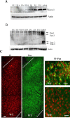
Figure 1.
Figure 1.
WAVE1 and pancortins are enriched in the adult mouse brain. A, B, Immunoblots for WAVE1 and pancortins in samples of embryonic (E12–E18), postnatal (P0–P15), and adult mouse cerebral cortex. C, Immunofluorescence imaging for pancortins (Pan; green) in cerebral cortex of adult WT mice and pancortin−/− mice (KO). A section of cerebral cortex with pancortin staining is shown. The dotted lines label the pial surface and the border between gray matter and white matter. Many cortical neurons in WT mice exhibit pancortin immunoreactivity, whereas no immunoreactivity is present in pancortin-deficient mice. The brain sections were counterstained with propidium iodide (PI; red) to label nuclei. A higher magnification of pancortin staining in cerebral cortex is shown. Scale bar, 20 μm.
Figure 2.
Figure 2.
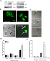
WAVE1 and pancortin-2 interact functionally to induce apoptosis. A, Immunoblot shows the overexpression of WAVE1–GFP and pancortin-2–GFP after 3 d of transfection in HEK293 cells. B–E, Coexpression of WAVE1–GFP and pancortin-2–GFP in HEK 293 cells (B, D) and primary cultured cortical neurons (C, E) caused cell death, whereas expression of either WAVE1–GFP or pancortin-2–GFP alone did not. Immunofluorescence images of HEK293 (B) or cortical neuronal (C) GFP+ cells overexpressing WAVE1 and pancortin-2 alone or in combination were shown. Expression of both WAVE1 and pancortin-2 results in cell shrinkage and the appearance of pyknotic nuclei. D, E, Values are the mean and SEM from three independent experiments. *p < 0.05.
Figure 3.
Figure 3.
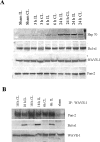
Focal brain ischemia induces the formation of a WAVE1/pancortin/Bcl-xL protein complex. A, Immunoblot for WAVE1, pancortin-2, Bcl-xL, and Hsp70 in samples of contralateral (CL) and ipsilateral (IL) cortical tissues lysates from sham or MCAO mice at different time points. B, The lysates were immunoprecipitated with WAVE1 antibodies and subjected to immunoblotting with antibodies to pancortin-2 and Bcl-xL. The blots were reprobed with WAVE1 antibodies to control for protein loading. IP, Immunoprecipitation.
Figure 4.
Figure 4.
Genetic deletion of pancortins ameliorates ischemic brain damage. A, B, Structure of the mouse pancortin gene with box representing exons (A, B, M1, M2, Y, Z1, Z2, Z3) and targeting vector that aimed to delete exon M2 (common to all isoforms) and exon Y. FRT, FLP recognition target; Pgk-Neo, phosphoglycerate kinase I promoter, driving the neomycin phospho-transferase gene. C, Representative PCR products for genotyping using two sets of primers (1 + 2 and 1 + 3). D, Immunoblot for pancortins in adult cerebral cortex lysates of WT and pancortin−/− mice.
Figure 5.
Figure 5.
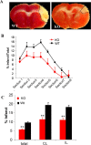
Cerebral infarct size is significantly reduced in pancortin−/− mice compared with WT mice. Pancortin−/− and WT were subjected to middle cerebral artery occlusion for 24 h. A, Representative TTC-stained brain sections (section 4) from WT and pancortin−/− mice. The infarct areas are circled by dotted lines. B, Quantification of infarct size at different rostro-caudal levels. C, Infarct volumes in WT and pancortin−/− mice expressed as percentages of total cortical volume, contralateral (CL) volume, or ipsilateral (IL) volume. **p < 0.01; n = 20 WT and 20 pancortin−/− mice. Error bars represent SEM.
Figure 6.
Figure 6.
Failure of WAVE1 to interact with Bcl-xL in pancortin−/− mice in response to focal brain ischemia. A, The amounts of WAVE1 and Bcl-xL are not altered in pancortin−/− mice. Immunoblot for WAVE1, Bcl-xL, and actin in cortical tissue samples from WT and pancortin−/− mice. Each lane is a sample from one mouse. B, C, The lysates from WT and pancortin−/− mice brains after 8 or 24 h MCAO were immunoprecipitated with WAVE1 antibodies and subjected to immunoblotting with antibodies to pancortin-2 and Bcl-xL. The blots were reprobed with WAVE1 antibodies to control for protein loading. D, Ischemia-induced binding of WAVE1 to Bcl-xL is disrupted in the cerebral cortex of pancortin−/− mice. Values are the mean and SEM (n = 6). *p < 0.01. CL, Contralateral; IL, ipsilateral; IP, immunoprecipitation.
Figure 7.
Figure 7.
The WAVE1/pancortin-2/Bcl-xL protein complex is associated with mitochondria. A, Immunoblot for Cox-1, Grp78, WAVE1, pancortin-2, Bcl-xL, and actin in cytosolic, mitochondrial, and microsomal cell fractions isolated from mouse cortical tissue by differential centrifugation. B, Immunoblot for WAVE1, Bcl-xL, and Cox-1 in mitochondrial fractions isolated from contralateral and ipsilateral cerebral cortical tissues from WT and pancortin−/− mice after 24 h of MCAO. C, Bax association with the mitochondria is suppressed after brain ischemia in pancortin−/− mice. A representative immunoblot of Bax in the mitochondrial fraction from both contralateral and ipsilateral cerebral cortex of WT and pancortin−/− mice is shown. Blots were re-probed with Cox-1 antibody to control for protein loading. D, Results of densitometric analysis of blots of samples from six different mice. Values are the mean and SEM (n = 6). *p < 0.01. E, The WAVE1/pancortin-2/Bcl-xL protein complex is present in high amounts in the mitochondrial fraction isolated from the ischemic cortex of WT mice but not in mitochondrial fractions from the ischemic cortex of pancortin−/− mice or nonischemic cortex (sham) of WT mice. The lysates of isolated mitochondria from ipsilateral brains were immunoprecipitated with WAVE1 antibodies and subjected to immunoblotting with antibodies to pancortin-2 and Bcl-xL. The blots were reprobed with WAVE1 antibodies to control for loading. F, A lesser percentage of mitochondria-associated Bax is bound with Bcl-xL in ischemic cortex in WT compared with pancortin−/− mice. Lysates of isolated mitochondria were immunoprecipitated with Bax antibody and immunoblotted with antibodies to Bcl-xL and Bax. Total Bcl-xL in mitochondria fraction was confirmed by separate Western blot. CL, Contralateral; IL, ipsilateral; IP, immunoprecipitation.
Figure 8.
Figure 8.
Cytochrome c release is suppressed after brain ischemia in pancortin−/− mice. A, Immunoblot analyses of cytochrome c (Cyto-c) in the cytosolic fraction and in the mitochondrial fraction from both contralateral (CL) and ipsilateral (IL) cerebral cortex of WT and pancortin−/− mice. Blots were reprobed with Erks (cytosol) and Cox-1 (mitochondria) antibodies to control for protein loading. B, Results of densitometric analysis of blots of samples from six different mice. Values are the mean and SD (n = 6). *p < 0.05 and **p < 0.01.

References

    1. Alto NM, Soderling J, Scott JD. Rab32 is an A-kinase anchoring protein and participates in mitochondrial dynamics. J Cell Biol. 2002;158:659–668. - PMC - PubMed
    1. Ando K, Nagano T, Nakamura A, Konno D, Yagi H, Sato M. Expression and characterization of disulfide bond use of oligomerized A2-pancortins: extracellular matrix constituents in the developing brain. Neuroscience. 2005;133:947–957. - PubMed
    1. Banzai Y, Miki H, Yamaguchi H, Takenawa T. Essential role of neural Wiskott-Aldrich syndrome protein in neurite extension in PC12 cells and rat hippocampal primary culture cells. J Biol Chem. 2000;275:11987–11992. - PubMed
    1. Basanez G, Sharpe JC, Galanis J, Brandt TB, Hardwick JM, Zimmerberg J. Bax-type apoptotic proteins porate pure lipid bilayers through a mechanism sensitive to intrinsic monolayer curvature. J Biol Chem. 2002;277:49360–49365. - PubMed
    1. Carnegie GK, Scott JD. A-kinase anchoring proteins and neuronal signaling mechanisms. Genes Dev. 2003;17:1557–1568. - PubMed

Publication types

MeSH terms

LinkOut - more resources