. 2007 Feb 14;27(7):1543-51.
doi: 10.1523/JNEUROSCI.3536-06.2007.

Activation of the extracellular signal-regulated kinase in the amygdala modulates pain perception

Affiliations

Activation of the extracellular signal-regulated kinase in the amygdala modulates pain perception

Yarimar Carrasquillo et al. J Neurosci. .

Abstract

The amygdala has been proposed to serve as a neural center for the modulation of pain perception. Numerous anatomical and behavioral studies demonstrate that exogenous manipulations of the amygdala (i.e., lesions, drug infusions) modulate behavioral responses to acute noxious stimuli; however, little is known about the endogenous molecular changes in the amygdala that contribute to alterations in nociceptive processing during persistent noxious stimuli that resemble pathological pain conditions. In the present study, we demonstrate that endogenous molecular changes in the amygdala play a crucial role in modulating long-lasting peripheral hypersensitivity associated with persistent inflammation and we further identify the extracellular signal-regulated kinase (ERK) as a molecular substrate underlying this behavioral sensitization. Using the formalin test as a mouse model of persistent inflammatory pain, we show that activation of ERK in the amygdala is both necessary for and sufficient to induce long-lasting peripheral hypersensitivity to tactile stimulation. Thus, blockade of inflammation-induced ERK activation in the amygdala significantly reduced long-lasting peripheral hypersensitivity associated with persistent inflammation, and pharmacological activation of ERK in the amygdala induced peripheral hypersensitivity in the absence of inflammation. Importantly, blockade of ERK activation in the amygdala did not affect responses to acute noxious stimuli in the absence of inflammation, indicating that modulation of nociceptive responses by amygdala ERK activation is specific to the persistent inflammatory state. Altogether, our results demonstrate a functional role of the ERK signaling cascade in the amygdala in inflammation-induced peripheral hypersensitivity.


Figures

Figure 1.
Figure 1.
Formalin-induced nociceptive behavior. a, Time course of formalin-induced spontaneous nociceptive behavior. Formalin injection induced triphasic spontaneous nociceptive behavior lasting for 2 h after injection (n = 9 animals for formalin treatment and 3 animals for saline treatment). b, Three hours after formalin injection, thermal latencies in the injected and noninjected hindpaw decreased to 51% (***p < 0.0001) and to 65% of baseline (**p = 0.0058), respectively, in formalin-injected but not saline-injected mice (n = 6 animals per treatment). c, Three hours after formalin injection, mechanical thresholds of the injected and noninjected paw decreased to 12% (**p = 0.0064, paired t test) and to 51% of baseline, respectively, (*p = 0.0205, paired t test) in formalin-injected but not saline-injected mice (n = 6 animals per treatment). Error bars indicate SEM.
Figure 2.
Figure 2.
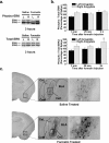
Formalin-induced amygdala ERK activation. a, Representative Western blots for phospho-ERK (top) and total-ERK (bottom) 3 h after formalin or saline paw injection. b, Densitometry analysis of bands corresponding to ERK1 (p44) and ERK2 (p42) show that ERK2 activation in the amygdala of formalin-injected animals is significantly greater than in saline-injected animals 3 h, but not 3 min or 25 min after formalin injection (n = 10 animals per treatment per time point; *p < 0.05, two-way ANOVA, Bonferroni's post hoc test). No significant ERK1 activation was observed. c, Anatomical distribution of formalin-induced ERK activation in the amygdala. Immunohistochemistry for phospho-ERK on coronal brain sections taken from animals 3 h after saline (top) or formalin (bottom) injection in the hindpaw. Formalin-induced ERK activation is localized to the laterocapsular subdivision of the central nucleus of the amygdala. Magnified views of the area delineated by the box are shown in the right panel. CeL, Lateral subdivision of central nucleus; CeC, capsular subdivision of central nucleus; CeM, medial subdivision of central nucleus; BLA, basolateral nucleus. Error bars indicate SEM.
Figure 3.
Figure 3.
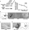
Inhibition of formalin-induced amygdala ERK activation by U0126. a, Representative Western blots for phospho-ERK and total-ERK 1 h after intra-amygdala drug infusion (left). U0126 was infused into the amygdala in doses ranging from 0.15 to 1.5 nmol in a total volume of 0.3 μl. Densitometry analysis of bands corresponding to ERK1 (p44) and ERK2 (p42) is shown in the right panel. U0126 inhibits phosphorylation of ERK in a dose-dependent manner (n = 3 animals; *p < 0.05, one-way ANOVA, Tukey's post hoc test). The asterisk indicates significant inhibition of ERK activation by U0126 compared with vehicle-treated animals. b, The first panel (left) is a diagram of a coronal section showing typical injection sites into the central nucleus of the amygdala (solid circles) or offsite injection sites into the perirhinal cortex (x). LA, Lateral nucleus; BLA, basolateral nucleus; PRh, perirhinal cortex. The second, third, and fourth panels are immunohistochemistry for phospho-ERK on coronal brain sections taken from animals after intra-amygdala or offsite drug infusion. U0126 (third panel), but not the control structural analog U0124 (second panel), inhibits formalin-induced amygdala ERK activation. Offsite infusion of U0126 (fourth panel) did not affect formalin-induced amygdala ERK activation. The tip of the injector is indicated by the arrow. c, Immunohistochemistry for phospho-ERK on a sagittal section. The arrows indicate the extent of ERK inhibition by U0126, with phospho-ERK immunopositive cells anterior and posterior to the injection site. d, Coinfusion of PDA with U0126; a diagram depicting the anatomical localization of different amygdala nuclei is shown in the right panel. Inhibition of PDA-induced ERK activation by U0126 is restricted to the CeA; U0126 extent of inhibition is delineated by the dotted line (left). Error bars indicate SEM.
Figure 4.
Figure 4.
Effects of intra-amygdala infusion of U0126 on nociceptive behavior. a, Spontaneous nociceptive responses to formalin were not significantly different in U0126- versus vehicle-injected mice (n = 8–10 animals per treatment). b, Baseline mechanical thresholds (in the absence of inflammation) are not affected by intra-amygdala infusion of vehicle (50% DMSO) or U0126 (1.5 nmol) (n = 3 mice per treatment). c, d, Five days after cannulation, animals were injected with 5% formalin into the hindpaw. Two hours after the paw injection, the MEK inhibitor U0126, the structural analog control compound U0124, or vehicle was infused into the amygdala. One hour after the amygdala injection, the effects of these treatments on thermal latencies (c) or mechanical thresholds (d) were analyzed. c, U0126 infusion into the amygdala did not affect formalin-induced thermal hypersensitivity (n = 4 animals per treatment). d, U0126 infusion significantly reduced formalin-induced mechanical hypersensitivity in both the injected and the noninjected hindpaw (n = 6–7 animals per treatment; p < 0.05, one-way ANOVA, Tukey's post hoc test, *p < 0.05; ***p < 0.0001). e, Offsite infusion of U0126 (1.5 nmol) did not affect formalin-induced mechanical hypersensitivity (n = 3 animals per treatment). Error bars indicate SEM.
Figure 5.
Figure 5.
PDA-induced ERK activation in the amygdala and effects on mechanical and thermal thresholds in the absence of inflammation. Five days after cannulation, mice received an infusion of the phorbol ester PDA (doses ranging from 45 to 450 pmol), or a coinfusion of PDA (120 pmol) plus U0126 (1.5 nmol) into the amygdala. In all cases, the total infusion volume was 0.3 μl. a, Extent of ERK activation by various doses of PDA. Infusion of PDA resulted in a robust dose-dependent increase in ERK activation in the amygdala. b, PDA (120 pmol) infusion into the amygdala did not affect baseline thermal thresholds in either the right or the left hindpaw (n = 6–7 animals per treatment). c, PDA (120 pmol) infusion into the amygdala decreased mechanical thresholds of both the right and the left hindpaw. PDA-induced behavior was blocked by coinfusion of PDA with U0126 (n = 5–6 animals; one-way ANOVA, Tukey's post hoc test, *p < 0.05). Error bars indicate SEM.

References

    1. Adamec RE, Blundell J, Burton P. Phosphorylated cyclic AMP response element binding protein expression induced in the periaqueductal gray by predator stress: its relationship to the stress experience, behavior and limbic neural plasticity. Prog Neuropsychopharmacol Biol Psychiatry. 2003;27:1243–1267. - PubMed
    1. Aggleton JP. The amygdala: a functional analysis. Ed 2. Oxford: Oxford UP; 2000.
    1. Aloisi AM, Porro CA, Cavazzuti M, Baraldi P, Carli G. “Mirror pain” in the formalin test: behavioral and 2-deoxyglucose studies. Pain. 1993;55:267–273. - PubMed
    1. Baker KB, Kim JJ. Amygdalar lateralization in fear conditioning: evidence for greater involvement of the right amygdala. Behav Neurosci. 2004;118:15–23. - PubMed
    1. Berman SM, Naliboff BD, Suyenobu B, Labus JS, Stains J, Bueller JA, Ruby K, Mayer EA. Sex differences in regional brain response to aversive pelvic visceral stimuli. Am J Physiol Regul Integr Comp Physiol. 2006;291:R268–R76. - PubMed

Publication types

MeSH terms