. 2007 Feb 14;27(7):1702-11.
doi: 10.1523/JNEUROSCI.5055-06.2007.

Identification of CRMP4 as a convergent regulator of axon outgrowth inhibition

Affiliations

Identification of CRMP4 as a convergent regulator of axon outgrowth inhibition

Yazan Z Alabed et al. J Neurosci. .

Abstract

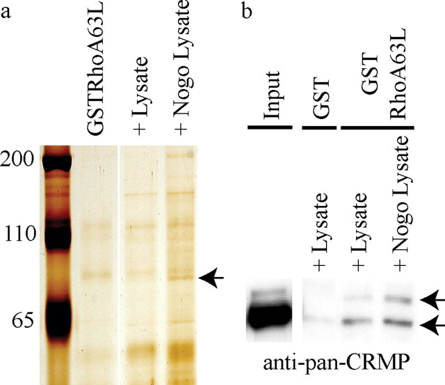
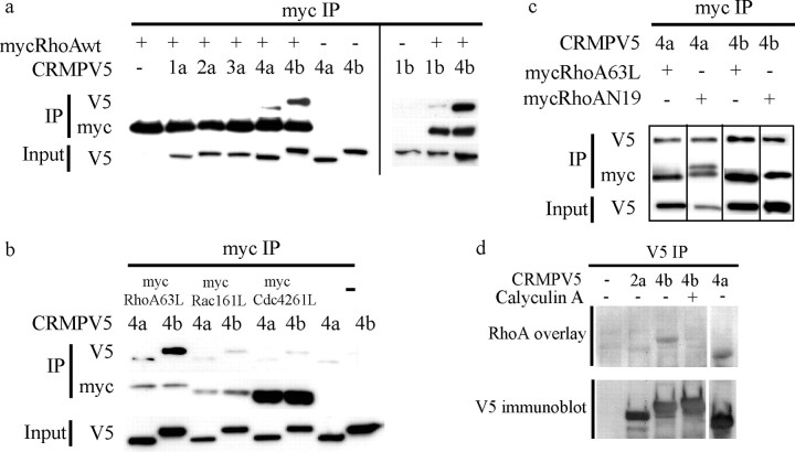
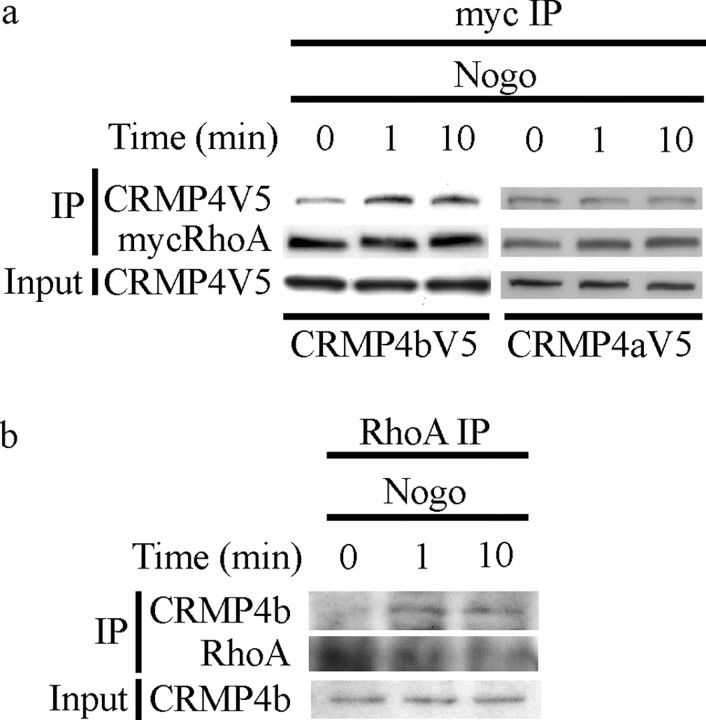
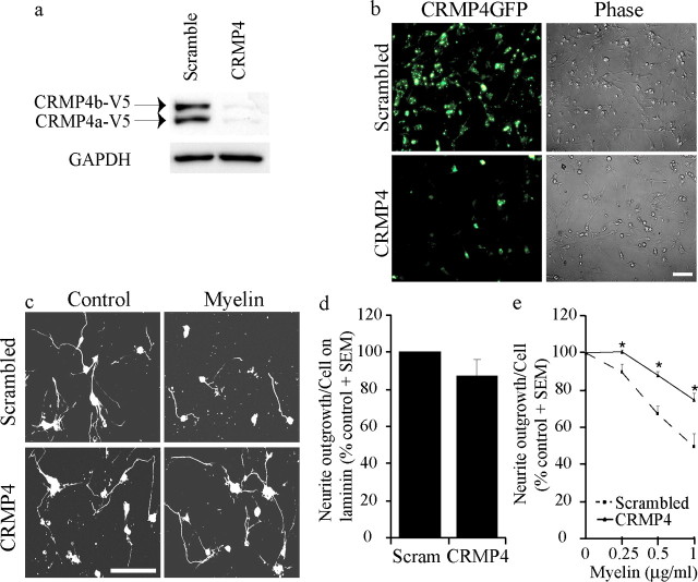
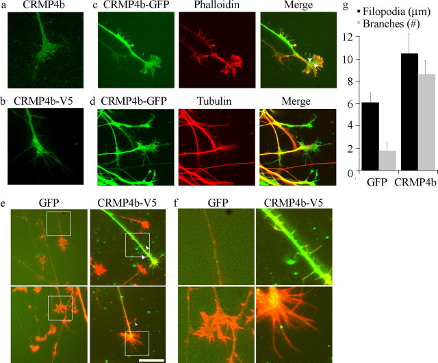
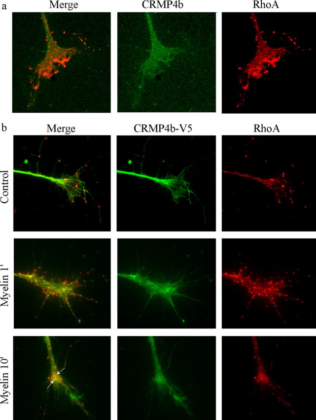
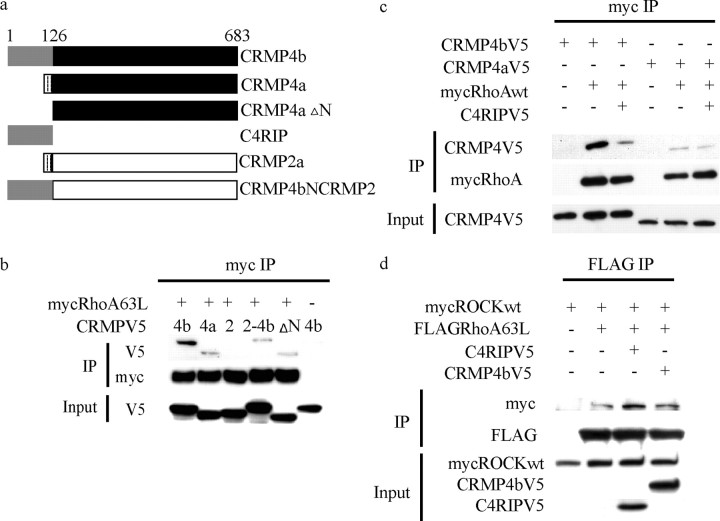
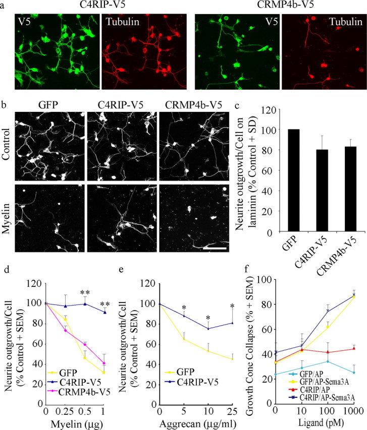
Myelin-associated inhibitors (MAIs) and chondroitin sulfate proteoglycans (CSPGs) contribute to failed regeneration after neuronal injury. MAIs and CSPGs stimulate intracellular signals including the activation of RhoA and Rho kinase to block axonal extension through targeted modifications to the cytoskeleton. RhoA and ROCK are promising targets for therapeutic intervention to promote CNS repair; however, their ubiquitous expression will limit the specificity of drugs targeted to these molecules. We have identified the cytosolic phosphoprotein CRMP4b (collapsin-response mediator protein 4b) as a protein that physically and functionally interacts with RhoA to mediate neurite outgrowth inhibition. Short interfering RNA-mediated knockdown of CRMP4 promotes neurite outgrowth on myelin substrates, indicating a critical role for CRMP4 in neurite outgrowth inhibition. Disruption of CRMP4b-RhoA binding with a competitive inhibitor attenuates neurite outgrowth inhibition on myelin and aggrecan substrates. Stimulation of neuronal growth cones with Nogo leads to colocalization of CRMP4b and RhoA at discrete regions within the actin-rich central and peripheral domains of the growth cone, indicative of a potential function in cytoskeletal rearrangements during neurite outgrowth inhibition. Together, these data indicate that a RhoA-CRMP4b complex forms in response to inhibitory challenges in the growth cone environment and regulates cytoskeletal dynamics at distinct sites necessary for axon outgrowth inhibition. Competitive inhibition of CRMP4b-RhoA binding suggests a novel, highly specific therapeutic avenue for promoting regeneration after CNS injury.


Figures

Figure 1.
Figure 1.
A novel RhoA–CRMP4 interaction is enhanced by Nogo-66. a, GSTRhoA63L pulldown from PC12 cells stimulated with AP (+ Lysate) or AP-Nogo-66 (+ Nogo Lysate). Precipitation of a 75 kDa protein identified as CRMP4b by tandem mass spectrometry is enhanced in the AP-Nogo-66-stimulated lysate (arrow). The protein runs slightly below a nonspecific protein from the GSTRhoA63L purification, which is visible in all lanes. The GSTRhoA63L lane represents beads that were not incubated with lysates. b, GST and GSTRhoA63L pull-downs from PC12 cell lysates immunoblotted with a pan-CRMP antibody. CRMPa (65 kDa) and CRMPb (75 kDa) bands are indicated in the PC12 pull-down lane (arrows).
Figure 2.
Figure 2.
The RhoA–CRMP4 interaction is highly specific, nucleotide independent, phospho dependent, and direct. a–c, HEK293T cells cotransfected with CRMP-V5 constructs and myc-tagged versions of wt and mutant Rho GTPases and subjected to myc immunoprecipitation. a, CRMP4 preferentially binds to RhoA. Data from two separate blots are separated by a vertical line. b, RhoA is the preferred binding partner for CRMP4. c, CRMP4 binding to RhoA is nucleotide independent. d, CRMP-V5 was immunoprecipitated from control or calyculin-treated HEK293T cells, separated by SDS-PAGE, and overlayed with recombinant RhoA. RhoA binds directly to CRMP4a and CRMP4b but fails to bind to phosphorylated CRMP4b. IP, Immunoprecipitation.
Figure 3.
Figure 3.
Nogo-P4 peptide enhances the interaction between CRMP4b and RhoA. a, b, PC12 cells transfected with myc-RhoA and CRMP4b-V5 or CRMP4a-V5; stimulated for 0, 1, or 10 min with Nogo-P4 peptide; and subjected to myc immunoprecipitation. b, P8 rat cerebellar cultures stimulated with Nogo-P4 peptide, subjected to RhoA immunoprecipitation, and analyzed for RhoA and CRMP4b. IP, Immunoprecipitation.
Figure 4.
Figure 4.
siRNA-mediated knockdown of CRMP4 expression promotes neurite outgrowth on myelin. a, HEK293T cells cotransfected with CRMP4-V5 and scrambled or CRMP4-targeted siRNA and analyzed by immunoblotting with anti-V5 antibody. GAPDH, Glyceraldehyde-3-phosphate dehydrogenase. b, Dissociated rat DRG neurons infected with HSVCRMP4b-GFP and transfected with scrambled or CRMP4 siRNA. Scale bar, 100 μm. c, βIII Tubulin-stained dissociated rat DRG neurons transfected with scrambled or CRMP4 siRNA and seeded on laminin (control) or myelin substrates for an 18 h neurite outgrowth assay. Scale bar, 100 μm. d, Quantitation of DRG outgrowth on control substrates with scrambled siRNA (Scram) or CRMP4 siRNA. e, Quantitation of DRG neurite outgrowth on myelin from neurons transfected with scrambled or CRMP4 siRNA. Values are normalized to baseline outgrowth on the control laminin substrate for each experiment. Determinations are from three experiments performed in duplicate. *p < 0.01 by Student's t test compared with scrambled siRNA.
Figure 5.
Figure 5.
CRMP4b overexpression promotes an actin-based filopodial phenotype in the neuronal growth cone and neurite. a, E13 chick DRG stained with an anti-CRMP4b antibody. b, E7 chick DRG neurons infected with HSV-CRMP4b-V5 and stained with anti-V5 antibody. c, d, E7 chick DRG neurons infected with HSV-CRMP4b-GFP and double stained with rhodamine-phalloidin (c) to label F-actin or anti-βIII tubulin antibody (d). e, E13 chick DRG neurons infected with HSV-GFP or HSV-CRMP4b-V5. GFP and CRMP4b-infected growth cones were stained with rhodamine-phalloidin (red). CRMP4b-V5 was stained with anti-V5 antibody (green). Scale bar, 10 μm. f, Magnification of boxed regions in d demonstrating enhanced neurite branching and filopodial extension in CRMP4b-V5-infected DRG neurons. g, Quantitation of number of branches per neurite and filopodial length in GFP- or CRMP4b-V5-infected DRG neurons.
Figure 6.
Figure 6.
CRMP4b and RhoA colocalize at discrete punctae during myelin-dependent growth cone collapse. a, b, Immunofluorescent detection of endogenous CRMP4b and RhoA (a) or CRMP4b-V5 and RhoA (b) in control or myelin-stimulated E13 chick DRG growth cones. Arrows in the merged panels indicate areas of CRMP4b-V5-RhoA colocalization.
Figure 7.
Figure 7.
The N-terminal domain of CRMP4b is sufficient for RhoA binding and disrupts full-length CRMP4b-RhoA binding when expressed as a recombinant fusion protein. a, Schematic of CRMP constructs generated to assess CRMP4 domains involved in RhoA binding. b, HEK293T cells cotransfected with myc-RhoA63L and CRMP-V5 constructs described in a and subjected to myc immunoprecipitation. c, HEK293T cells cotransfected with myc-RhoAWT, CRMP4-V5, and C4RIP-V5 to assess the effect of C4RIP on full-length CRMP4 binding to RhoA. d, HEK293T cells cotransfected with FLAG-RhoA63L, myc-ROCK, and C4RIP-V5 to assess the effect of C4RIP on RhoA binding to ROCK. IP, Immunoprecipitation.
Figure 8.
Figure 8.
C4RIP-V5 attenuates myelin inhibition. a, E13-dissociated chick DRG neurons infected with HSV-C4RIP-V5 or HSV-CRMP4b-V5 and double stained with anti-V5 and anti-βIII tubulin antibodies to validate neuronal infection. b, E13-dissociated chick DRG neurons infected with C4RIP-V5 or CRMP4b-V5 plated on laminin (control) or myelin substrates and stained with anti-βIII tubulin to visualize neurite outgrowth. c–e, Quantitation of neurite outgrowth from GFP-, C4RIP-, or CRMP4b-infected neurons on laminin (c), myelin (d), or aggrecan (e) substrates. In c, neurite outgrowth per cell is normalized to outgrowth in GFP-infected neurons for each experiment (100%). In d and e, neurite outgrowth is normalized to growth on the laminin substrate for each dose curve (100%). Determinations are from four separate experiments. Scale bar, 100 μm. *p < 0.05 and **p < 0.01 by Student's t test compared with GFP. f, Quantitation of growth cone collapse in E8 chick DRG neurons infected with HSV-GFP or HSV-C4RIP and stimulated for 20 min with control AP ligand or AP-Sema3A ligand. Determinations are from three separate experiments performed in duplicate.

References

    1. Alabed YZ, Grados-Munro E, Ferraro GB, Hsieh S, Fournier AE. Neuronal responses to myelin are mediated by ROCK. J Neurochem. 2006;96:1616–1625. - PubMed
    1. Arimura N, Menager C, Kawano Y, Yoshimura T, Kawabata S, Hattori A, Fukata Y, Amano M, Goshima Y, Inagaki M, Morone N, Usukura J, Kaibuchi K. Phosphorylation by Rho kinase regulates CRMP-2 activity in growth cones. Mol Cell Biol. 2005;25:9973–9984. - PMC - PubMed
    1. Arthur WT, Ellerbroek SM, Der CJ, Burridge K, Wennerberg K. XPLN, a guanine nucleotide exchange factor for RhoA and RhoB, but not RhoC. J Biol Chem. 2002;277:42964–42972. - PubMed
    1. Bishop AL, Hall A. Rho GTPases and their effector proteins. Biochem J. 2000;348:241–255. - PMC - PubMed
    1. Borisoff JF, Chan CC, Hiebert GW, Oschipok L, Robertson GS, Zamboni R, Steeves JD, Tetzlaff W. Suppression of Rho-kinase activity promotes axonal growth on inhibitory CNS substrates. Mol Cell Neurosci. 2003;22:405–416. - PubMed

Publication types

MeSH terms

LinkOut - more resources