. 2007 Feb 20;104(8):2631-6.
doi: 10.1073/pnas.0606822104. Epub 2007 Feb 14.

Bilayer deformation by the Kv channel voltage sensor domain revealed by self-assembly simulations

Affiliations

Bilayer deformation by the Kv channel voltage sensor domain revealed by self-assembly simulations

Peter J Bond et al. Proc Natl Acad Sci U S A. .

Abstract

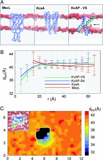
Coarse-grained molecular dynamics simulations are used to explore the interaction with a phospholipid bilayer of the voltage sensor (VS) domain and the S4 helix from the archaebacterial voltage-gated potassium (Kv) channel KvAP. Multiple 2-mus self-assembly simulations reveal that the isolated S4 helix may adopt either interfacial or transmembrane (TM) locations with approximately equal probability. In the TM state, the insertion of the voltage-sensing region of S4 is facilitated via local bilayer deformation that, combined with side chain "snorkeling," enables its Arg side chains to interact with lipid headgroups and water. Multiple 0.2-mus self-assembly simulations of the VS domain are also performed, along with simulations of MscL and KcsA, to permit comparison with more "canonical" integral membrane protein structures. All three stably adopt a TM orientation within a bilayer. For MscL and KcsA, there is no significant bilayer deformation. In contrast, for the VS, there is considerable local deformation, which is again primarily due to the lipid-exposed S4. It is shown that for both the VS and isolated S4 helix, the positively charged side chains of S4 are accommodated within the membrane through a combination of stabilizing interactions with lipid glycerol and headgroup regions, water, and anionic side chains. Our results support the possibility that bilayer deformation around key gating charge residues in Kv channels may result in "focusing" of the electrostatic field, and indicate that, when considering competing models of voltage-sensing, it is essential to consider the dynamics and structure of not only the protein but also of the local lipid environment.


Conflict of interest statement

The authors declare no conflict of interest.

Figures

Fig. 1.
Fig. 1.
The VS domain (PDB ID 1ORS) from KvAP. (A) Atomistic structure of the VS, showing non-H atoms only in space-filling format with the S4 helix in (pale) blue and the Arg side chains of S4 in deep blue. (B) CG model of the VS, shown in space-filling format with the S4 helix in pale blue and the Arg residues of S4 in deep blue.
Fig. 2.
Fig. 2.
Interactions of the S4 helix with a PC bilayer. (A) CG model of the S4 helix (residues 115–153) from KvAP showing the location of the side chain particles for basic (blue), acidic (red), and polar (pink) residues. This model, in a box of 256 DPPC molecules and 3,150 water particles, was the starting point for five CG-MD simulations, each of duration 2 μs. (B) Snapshots from the end of the S4 CG-MD simulations showing the S4 helix located at the lipid/water interface. (C) Snapshots from the end of the S4 CG-MD simulations showing the S4 helix inserted in the bilayer and switching between an extended helix and kinked helix conformation. The glycerol backbone particles are shown as green spheres.
Fig. 3.
Fig. 3.
CG-MD simulations of membrane protein insertion into a lipid (DPPC) bilayer. (A) Three membrane proteins inserted in a bilayer via CG-MD self-assembly simulations: MscL, KcsA, and the KvAP VS domain. In each case, the protein is shown as a Cα trace, and the glycerol backbone particles of the bilayer are represented as red spheres. For KvAP, the S4 helix is highlighted in green, whereas significant bilayer deformation is indicated by a horizontal arrow. (B) Local deformation of the lipid bilayer measured as the average distance (±SD) between upper and lower P particles (dPP) vs. the distance of the particles in the xy plane from the centre of mass of the corresponding protein (r). The curve for S4 corresponds to analysis of a simulation in which S4 adopted a TM orientation (similar results were obtained from the other such simulations). (C) Local deformation of the lipid bilayer around the KvAP VS measured as the average distance between upper and lower P particles (dPP) as a function of position in the xy plane. The central black object corresponds to the area occupied by the protein.
Fig. 4.
Fig. 4.
Analysis of S4/lipid and S4/water contacts in S4, averaged over all bilayer-inserted S4 and VS simulations. (A) Contacts (interparticle distance <6 Å) between the basic side chains of the isolated S4 helix and the water (W, red), PO4 (P, black), and glycerol (G, blue) particles. For each side chain, the mean (±SD) number of contacts over the second half of the simulation is shown. (B) Contacts between the basic side chains of the S4 helix within the KvAP VS domain and: water (W, red), PO4 (P, black), glycerol (G, blue), and anionic side chain (A, green) particles. For each side chain, the mean (±SD) number of contacts over the second half of the simulation is shown. (C) Total contacts (with all particles) of the basic side chains of S4, and of S4 within the KvAP domain (black).

References

    1. Hille B. Ionic Channels of Excitable Membranes. Sunderland, MA: Sinauer; 2001.
    1. Bezanilla F. Physiol Rev. 2000;80:555–592. - PubMed
    1. Yellen G. Nature. 2002;419:35–42. - PubMed
    1. Swartz KJ. Nat Rev Neurosci. 2004;5:905–916. - PubMed
    1. Jiang Y, Lee A, Chen J, Ruta V, Cadene M, Chait BT, Mackinnon R. Nature. 2003;423:33–41. - PubMed

Publication types

LinkOut - more resources