. 2007 Feb 15;445(7129):732-7.
doi: 10.1038/nature05580.

Structural definition of a conserved neutralization epitope on HIV-1 gp120

Affiliations

Structural definition of a conserved neutralization epitope on HIV-1 gp120

Tongqing Zhou et al. Nature. .

Abstract

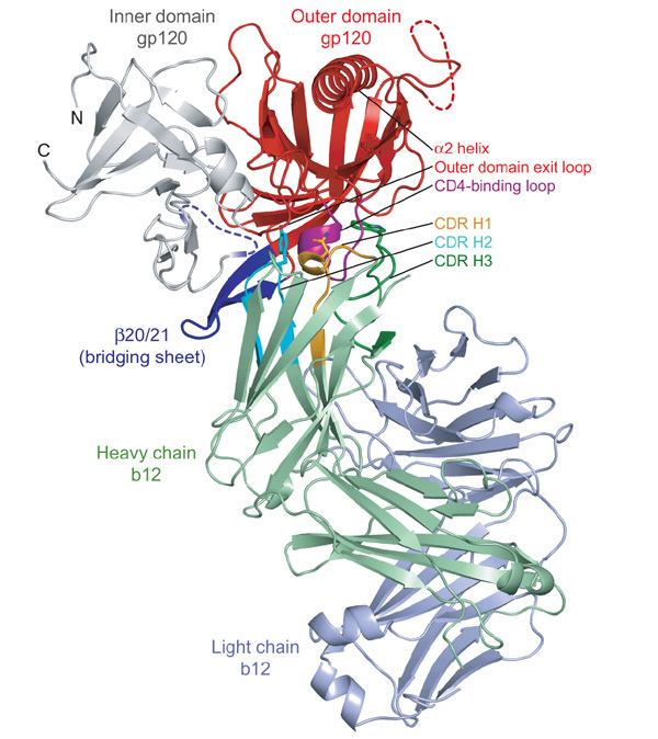
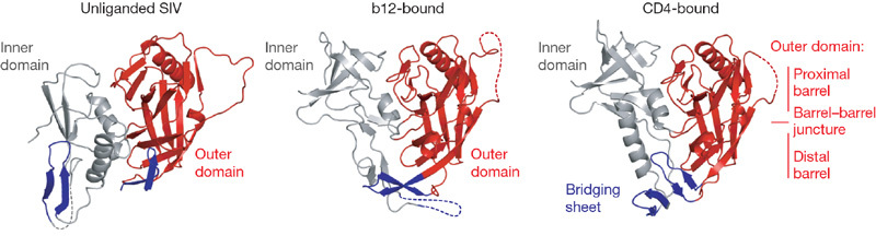
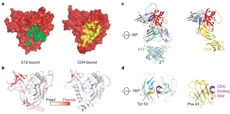
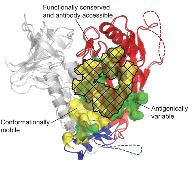
The remarkable diversity, glycosylation and conformational flexibility of the human immunodeficiency virus type 1 (HIV-1) envelope (Env), including substantial rearrangement of the gp120 glycoprotein upon binding the CD4 receptor, allow it to evade antibody-mediated neutralization. Despite this complexity, the HIV-1 Env must retain conserved determinants that mediate CD4 binding. To evaluate how these determinants might provide opportunities for antibody recognition, we created variants of gp120 stabilized in the CD4-bound state, assessed binding of CD4 and of receptor-binding-site antibodies, and determined the structure at 2.3 A resolution of the broadly neutralizing antibody b12 in complex with gp120. b12 binds to a conformationally invariant surface that overlaps a distinct subset of the CD4-binding site. This surface is involved in the metastable attachment of CD4, before the gp120 rearrangement required for stable engagement. A site of vulnerability, related to a functional requirement for efficient association with CD4, can therefore be targeted by antibody to neutralize HIV-1.


Conflict of interest statement

Coordinates and structure factors have been deposited in the Protein Data Bank and may be obtained from the authors (accession codes 2nxy–2ny6 for the nine variant gp120 molecules with CD4 and 17b; accession code 2ny7 for the b12–gp120 complex). Reprints and permissions information is available at www.nature.com/reprints. The authors declare no competing financial interests.

Figures

Figure 1
Figure 1. Structure of b12 in complex with an HIV-1 gp120 core.
Polypeptide chains are depicted in ribbon representation, with disordered regions as dashed lines. The gp120 inner domain is grey, and the outer domain is red, except for the CD4-binding loop, which is purple. The strands and associated loops, which in the CD4-bound conformation correspond to the bridging sheet, are blue. The b12 light chain is blue–grey and the b12 heavy chain is green, with associated CDRs highlighted in orange (H1), cyan (H2) and dark green (H3). The heavy-chain dominance of the binding interaction is apparent, with the nearest light-chain approach separated by ∼10 Å from gp120. Heavy-chain-only interactions are rare, although heavy-chain interactions predominate in a number of viral Env–antibody complexes, including those from SARS coronavirus and influenza virus haemagglutinin. Three b12 residues (Asn 31, Tyr 53 and Trp 100) from each of the heavy-chain CDRs are depicted in stick representation. Together, these three residues combine to form ∼40% of the b12 contact surface. They can be seen gripping the CD4-binding loop, the central focus of the b12 interaction with gp120.
Figure 2
Figure 2. Conformational states of gp120.
The unliganded, b12- and CD4-bound conformations of gp120 are depicted, with polypeptide in ribbon representation and disordered regions as dashed lines. Inner domains are grey, outer domains are red and regions that in the CD4-bound state correspond to the bridging sheet are blue. Both b12- and CD4-bound conformations are of the Ds12 F123 variant of HIV-1, whereas the unliganded structure is of simian immunodeficiency virus (SIV). Comparison of these three gp120 conformations highlights not only the structural plasticity of the inner domain and bridging sheet, but also the conformational stability of the outer domain.
Figure 3
Figure 3. b12 and CD4 recognition of gp120.
The orientation of gp120 in a and b is the same as in Fig. 2, with c and d rotated about a horizontal axis by 90° and 180°, respectively. a, Molecular surface of gp120 in red, with the b12-contact surface in green (left) and the CD4-contact surface in yellow (right). b, Ribbon diagram of the b12- and CD4-bound gp120 coloured according to the atomic mobility of the polypeptide, with white for fixed and red for flexible. In the b12-induced conformation, only the outer domain is fixed by interaction with antibody, with the average atomic mobility of the outer domain about one-half that of the inner domain. By contrast, CD4 fixes the entire core, resulting in outer and inner domains of similar overall atomic mobility. c, Comparison of b12 and CD4 angles of attachment. The polypeptide chains are depicted in ribbon representation, and coloured according to Fig. 1, with CD4 in yellow. The similarity in angle of attachment as well as the binding focus of b12 on the outer domain (red) are evident in this orientation. d, The CD4-binding loop of gp120 (purple) is shown with b12 (left) and CD4 (right). There are parallels between several key CD4 contact residues and those of b12. For example, Phe 43 of CD4 (shown in stick model) inserts into a critical juncture at the nexus of the inner, outer and bridging sheet regions of gp120, whereas Tyr 53 of b12 (also shown in stick model) inserts at a similar position, although displaced by ∼3 Å.
Figure 4
Figure 4. Structural definition of a conformationally invariant, antibody-accessible portion of the CD4-binding site.
The b12- and CD4-bound conformations of gp120 are shown in ribbon representation, after superposition of outer domains (red). A semitransparent molecular surface shows the contact surfaces of b12 (green) and CD4 (yellow). Subsets of these surfaces, corresponding to regions of conformational flexibility (for example, of the inner domain (grey) or bridging sheet (blue)), are delineated, as are regions of b12 contact outside of the conserved CD4-binding site. As can be seen, functional analysis serves to transcend the particulars of b12 binding, whereas antibody defines accessibility. Although we have formally shown only the b12 contact surface to be accessible in the context of a functional viral spike, the highly effective neutralization of D1D2-Igαtp and the kinetics of its association with both core and OD1 variants of gp120 suggest that the CD4-binding surface on the outer domain is accessible.

Comment in

References

    1. Korber B, et al. Timing the ancestor of the HIV-1 pandemic strain. Science. 2000;288:1789–1796. doi: 10.1126/science.288.5472.1789. - DOI - PubMed
    1. Joint United National Programme on HIV/AIDS. 2006 Report on the global AIDS epidemic. 〈http://www.aids.org/en/HIVdata/2006GlobalReport/〉 (2006)
    1. Weiss RA, et al. Neutralization of human T-lymphotropic virus type III by sera of AIDS and AIDS-risk patients. Nature. 1985;316:69–72. doi: 10.1038/316069a0. - DOI - PubMed
    1. Wyatt R, Sodroski J. The HIV-1 envelope glycoproteins: fusogens, antigens and immunogens. Science. 1998;280:1884–1888. doi: 10.1126/science.280.5371.1884. - DOI - PubMed
    1. Parren PW, Moore JP, Burton DR, Sattentua QJ. The neutralizing antibody response to HIV-1: viral evasion and escape from humoral immunity. AIDS. 1999;13:S137–S162. - PubMed

Publication types

MeSH terms