. 2007 Feb 14;2(2):e204.
doi: 10.1371/journal.pone.0000204.

Ancient origin of the new developmental superfamily DANGER

Affiliations

Ancient origin of the new developmental superfamily DANGER

Nikolas Nikolaidis et al. PLoS One. .

Abstract

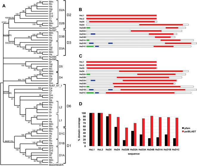
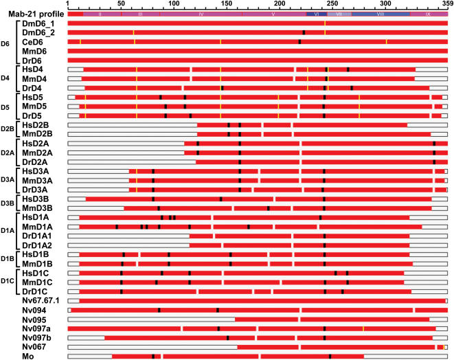
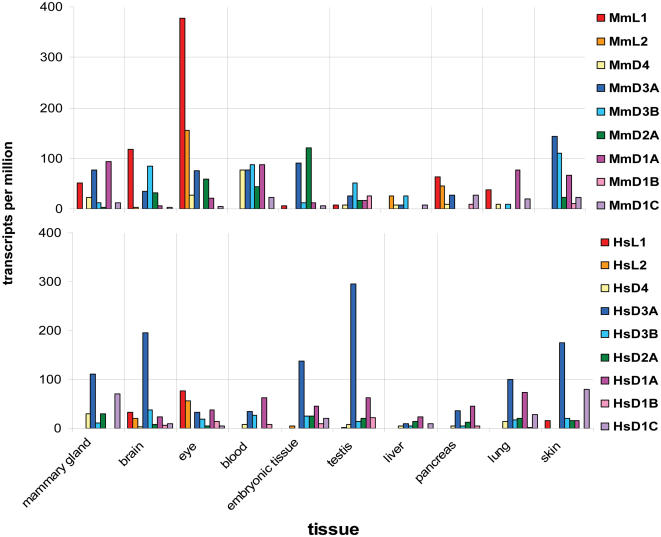
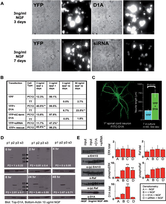
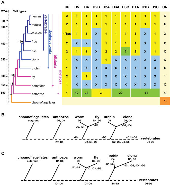
Developmental proteins play a pivotal role in the origin of animal complexity and diversity. We report here the identification of a highly divergent developmental protein superfamily (DANGER), which originated before the emergence of animals (approximately 850 million years ago) and experienced major expansion-contraction events during metazoan evolution. Sequence analysis demonstrates that DANGER proteins diverged via multiple mechanisms, including amino acid substitution, intron gain and/or loss, and recombination. Divergence for DANGER proteins is substantially greater than for the prototypic member of the superfamily (Mab-21 family) and other developmental protein families (e.g., WNT proteins). DANGER proteins are widely expressed and display species-dependent tissue expression patterns, with many members having roles in development. DANGER1A, which regulates the inositol trisphosphate receptor, promotes the differentiation and outgrowth of neuronal processes. Regulation of development may be a universal function of DANGER family members. This family provides a model system to investigate how rapid protein divergence contributes to morphological complexity.


Conflict of interest statement

Competing Interests: The authors have declared that no competing interests exist.

Figures

Figure 1
Figure 1. DANGER proteins originated early in metazoan evolution and encode a highly divergent MAB-21 domain.
(A) Neighbor-joining (NJ) consensus tree of the DANGER superfamily defines orthologous relationships among DANGER sequences (D1–D6) from vertebrates (Dr, Danio rerio; Xl, Xenopus laevis; Xt, Xenopus tropicalis; Gg, Gallus gallus; Rn, Rattus norvegicus; Mm, Mus musculus; Hs, Homo sapiens), invertebrates (Ci, Ciona intestinalis; Sp, Strongylocentrotus purpuratus; Dm, Drosophila melanogaster; Ce, Caenorhabditis elegans; Cb, Caenorhabditis briggsae, Nv, Nematostella vectensis), and the choanoflagellate (Mo, Monosiga ovata). Numbers at branches are bootstrap values from the NJ analysis. Bootstrap and quartet puzzling support values from maximum parsimony (MP) and maximum likelihood (TREE-PUZZLE) analyses, respectively, are given in parentheses. (B) Domain prediction analysis using the Pfam Mab-21 profile, containing four sequences, identifies a partial Mab-21 domain (showed as red boxes) in the human DANGER (HsD1-5) proteins. Predicted signal peptides and transmembrane regions are depicted by green and blue boxes, respectively. (C) Domain prediction analysis, using a Mab-21 profile generated by psi-BLAST containing 63 animal sequences, results in Mab-21 domain extension in all human DANGER proteins. (D) Quantification of Mab-21 domain coverage as predicted by the Pfam and the psiBLAST-generated Mab-21 profiles in all human DANGER proteins. Similar Mab-21 domain extension is observed between orthologous DANGER sequences from other species.
Figure 2
Figure 2. Patterns of sequence evolution along the Mab-21 domain.
(A) Graphical representation of the p-distances calculated along the multiple sequence alignment of representative DANGER proteins with a sliding window of 100 amino acids and a step of 50 amino acids. Bars represent standard errors of mean p-distance values. (B) Multiple sequence alignment of representative DANGER sequences (D1–D6) in block format. Microdomains II to IX are colored according to the mean value of the proportion of amino acid differences (p-distance) among all sequences. Intron positions are mapped as yellow arrows. (B) Graphical representation of the p-distances for each microdomain. Bars represent standard errors of mean p-distance values. (D) Pattern of sequence conservation (logo) along the three most conserved Mab-21 microdomains (VI–VIII).
Figure 3
Figure 3. Evolutionary patterns of the Mab-21 domain sequence.
Rps-BLAST pairwise alignments between the psi-BLAST generated Mab-21 profile and DANGER sequences. Comparison of insertions (in profile sequence; black boxes) and deletions (in profile sequence; white boxes), and correspondence of indels (>3 amino acids) with intron positions (yellow boxes). For comparison, all DANGER sequences are mapped onto the Mab-21 profile sequence according to pairwise alignment coordinates. The microdomains II–VIII are also shown. Species abbreviations are as in Figure 1A.
Figure 4
Figure 4. Analysis of expressed sequence tags (ESTs) show that mouse and human DANGER sequences exhibit different transcriptional patterns.
Upper: Plot depicting the expression levels of the mouse DANGER sequences in different tissues. Lower: Plot depicting the expression levels of the human DANGER sequences in different tissues. (Additional information and the Unigene identification numbers for each gene used in this analysis can be found in Figure S13).
Figure 5
Figure 5. DANGER1A increases neurite outgrowth in response to NGF signaling.
(A) Top: 40× Phase picture and 485 nm emission of PC12 cells transfected with YFP-alone or YFP+MYC-tagged D1A stimulated with NGF (3 ng/ml 3 days). PC12 cells expressing D1A have increased neurite outgrowth. Bottom: Phase picture and 485 nm emission of PC12 cells transfected with YFP-alone or YFP+siRNA against rat D1A stimulated with NGF (3 ng/ml 7 days). PC12 cells depleted of endogenous D1A using siRNA have decreased neurite outgrowth. (B) Quantification of neurite outgrowth in PC12 and TT cells. (100 cells counted in 3 independent experiments) (C) Left: Immunofluorescent staining of mouse primary spinal cord neurons with Alexa488-conjugated DANGER1A antibody. Inset: Quantification of spinal cord neurite length in neurons overexpressing YFP alone or YFP+D1A. (D) Western analysis of D1A expression in the p1 (nuclear), p2 (heavy ER and mitochondria), p3(light ER and vesicles), or s3 (cytosol) cell fractions over a time course of 48 h stimulation with NFG (10 ng/ml). These individual blots were developed simultaneously on the same film. The numbers inside the blots quantify the change in D1A distribution over time course (Error: SEM; p<0.01 n = 4). (E) Right: Western analysis of the MAP-kinase proteins ERK1/2, phospho-ERK1/2, Raf, phospho-Raf, and D1A levels in PC12 cells transfected with either YFP, YFP+D1A, or YFP+siRNA D1A. Cells were treated with vehicle (water), or 3 ng/ml NGF for 48 hours. Left: Quantification of changes in total and phospho-ERK1/2 and total D1A by scanning densitometry. All values are expressed as fold-change vs. control; ERK-(Error: SEM; p<0.05 n = 3); D1A-(Error: SEM; p<0.01 n = 3)
Figure 6
Figure 6. Distribution and evolution of DANGER genes in metazoa.
(A) The tree on the left summarizes the phylogenetic relationships of the informative species used. Numbers at nodes represent cell types adopted from . Yellow boxes indicate the presence of a DANGER gene in a particular taxon. Numbers correspond to the number of genes found in each taxon. “X” indicates the absence of a DANGER gene in a particular taxon (blue boxes). Uncertain orthologous relationships are indicated by question marks (green boxes). “UN” indicates the unclassified DANGER proteins or the ones that could not be unconditionally assigned to a specific group. The choanoflagellate sequence from M. ovata assumes outgroup position in all phylogenetic analyses. Divergence times in million years ago (MYA) were taken from references , . (B–C) Two alternative evolutionary scenarios explain the evolution of DANGER families in metazoa. Both scenarios presume that the genome sequence of the vertebrate ancestor encoded at least six DANGER lineages, namely D1–D6. (B) According to the first scenario, anthozoa contain two (D2, D6) of the six vertebrate DANGER lineages, while the remaining DANGER lineages have evolved by repeated cycles of gene birth (+) and death (−). (C) In the second scenario anthozoa species contain sequences orthologous to all vertebrate DANGER groups. This scenario presupposes that in ecdysozoa, hemichordates, and urochordates four DANGER groups were lost. Dotted lines correspond to ancestral stages. The order of events is presented according to the species tree.

References

    1. Carroll SB, Grenier JK, Weatherbee SD. From DNA to Diversity: Molecular Genetics and the Evolution of Animal Design. Malden, MA: Blackwell Publishers; 2004.
    1. Davidson EH, Erwin DH. Gene regulatory networks and the evolution of animal body plans. Science. 2006;311:796–800. - PubMed
    1. Avise JC. Phylogeography: The History and Formation of Species. Cambridge, MA: Harvard University Press; 2000.
    1. van Rossum DB, Patterson RL, Cheung KH, Barrow RK, Syrovatkina V, et al. DANGER, a novel regulatory protein of inositol 1,4,5-trisphosphate-receptor activity. J Biol Chem. 2006;281:37111–37116. - PubMed
    1. Chow KL, Hall DH, Emmons SW. The mab-21 gene of Caenorhabditis elegans encodes a novel protein required for choice of alternate cell fates. Development. 1995;121:3615–3626. - PubMed

Publication types

MeSH terms