. 2007 Jul;212(1):207-14.
doi: 10.1002/jcp.21021.

Oscillating fluid flow activation of gap junction hemichannels induces ATP release from MLO-Y4 osteocytes

Affiliations

Oscillating fluid flow activation of gap junction hemichannels induces ATP release from MLO-Y4 osteocytes

Damian C Genetos et al. J Cell Physiol. 2007 Jul.

Abstract

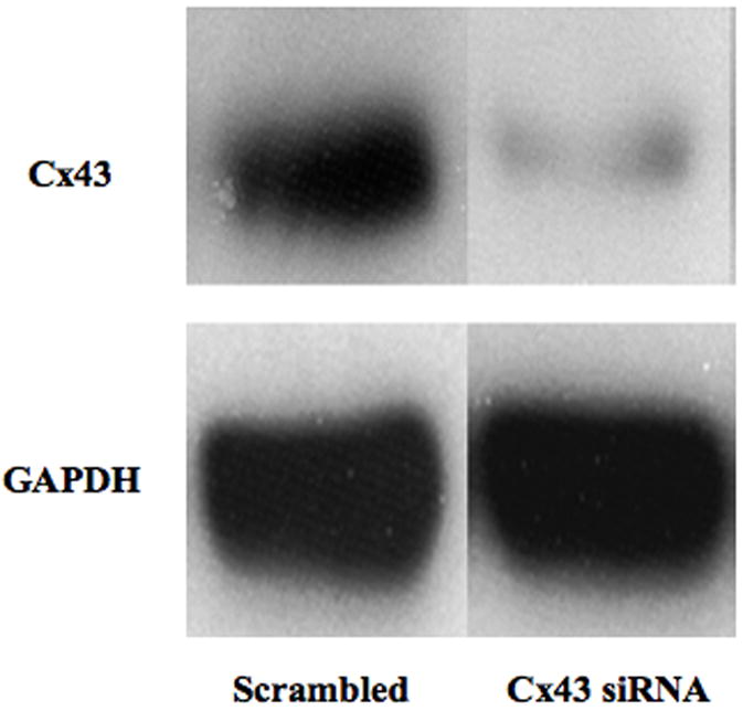
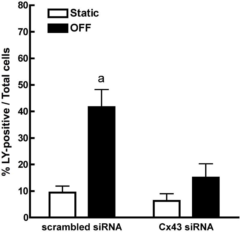
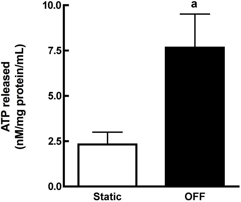
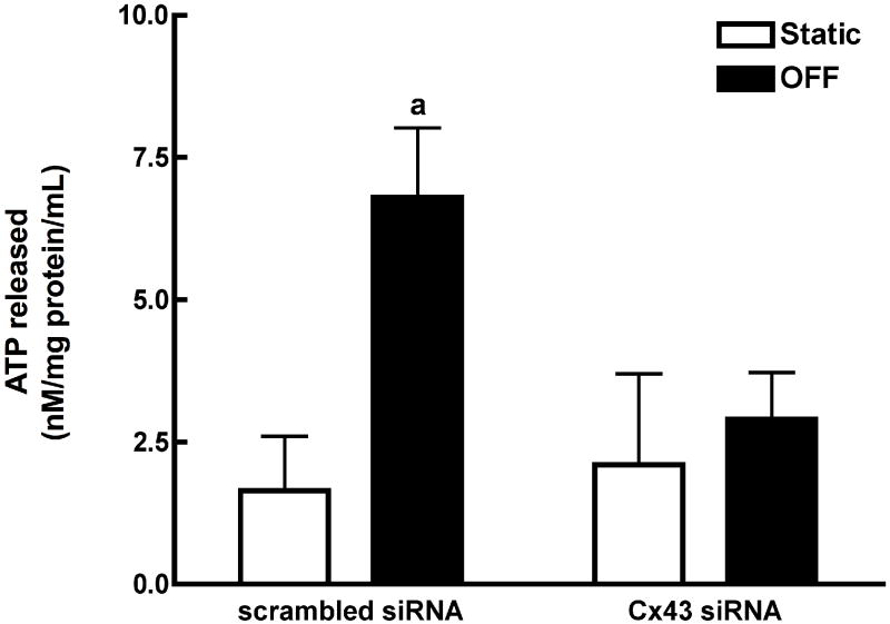
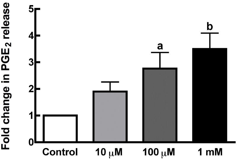
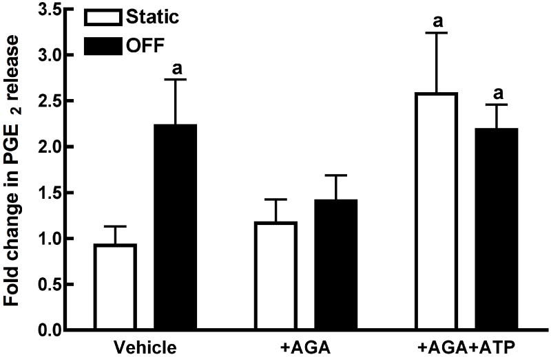
Mechanical loads are required for optimal bone mass. One mechanism whereby mechanical loads are transduced into localized cellular signals is strain-induced fluid flow through lacunae and canaliculi of bone. Gap junctions (GJs) between osteocytes and osteoblasts provides a mechanism whereby flow-induced signals are detected by osteocytes and transduced to osteoblasts. We have demonstrated the importance of GJ and gap junctional intercellular communication (GJIC) in intracellular calcium and prostaglandin E(2) (PGE(2)) increases in response to flow. Unapposed connexons, or hemichannels, are themselves functional and may constitute a novel mechanotransduction mechanism. Using MC3T3-E1 osteoblasts and MLO-Y4 osteocytes, we examined the time course and mechanism of hemichannel activation in response to fluid flow, the composition of the hemichannels, and the role of hemichannels in flow-induced ATP release. We demonstrate that fluid flow activates hemichannels in MLO-Y4, but not MC3T3-E1, through a mechanism involving protein kinase C, which induces ATP and PGE(2) release.


Figures

Figure 1
Figure 1
Lucifer Yellow dye uptake through hemichannels occurs in MLO-Y4 osteocytes but not MC3T3-E1 osteoblasts. (A) Quantitation of Lucifer Yellow dye uptake in response to oscillatory fluid flow in MLO-Y4 osteocytes and MC3T3-E1 osteoblasts. MLO-Y4 osteocytes exposed to oscillatory fluid flow take up extracellular Lucifer Yellow that is inhibited in the presence of the hemichannel antagonist AGA. In contrast, MC3T3-E1 osteoblasts exposed to oscillatory fluid flow do not take up extracellular Lucifer Yellow, and the hemichannel antagonist AGA has no effect on dye uptake. Data are presented as the mean percentage of cells staining positively for Lucifer Yellow divided by the total number of cells, visualized by Hoechst 33258, within a field of view. Data were obtained from 4 independent experiments. (a: p < 0.01 compared to static MLO-Y4; b: p < 0.01 compared to MLO-Y4 exposed to oscillatory fluid flow). (B) Quantitation of Lucifer Yellow dye uptake in response to 5mM EGTA treatment in static MLO-Y4 osteocytes and MC3T3-E1 osteoblasts. Lucifer yellow dye uptake and hemichannel activation in response to oscillatory fluid flow is mimicked in static cells treated with 5mM EGTA. MLO-Y4 osteocytes treated with 5mM EGTA take up extracellular Lucifer Yellow that is inhibited in the presence of the hemichannel antagonist AGA. MC3T3-E1 osteoblasts treated with 5mM EGTA do not take up extracellular Lucifer Yellow, and the hemichannel antagonist AGA has no effect on dye uptake. Data are presented as the mean percentage of cells staining positively for Lucifer Yellow divided by the total number of cells, visualized by brightfield imaging. Data were obtained from three independent experiments. (a: p < 0.01 compared to static MLO-Y4; b: p < 0.01 compared to MLO-Y4+EGTA).
Figure 1
Figure 1
Lucifer Yellow dye uptake through hemichannels occurs in MLO-Y4 osteocytes but not MC3T3-E1 osteoblasts. (A) Quantitation of Lucifer Yellow dye uptake in response to oscillatory fluid flow in MLO-Y4 osteocytes and MC3T3-E1 osteoblasts. MLO-Y4 osteocytes exposed to oscillatory fluid flow take up extracellular Lucifer Yellow that is inhibited in the presence of the hemichannel antagonist AGA. In contrast, MC3T3-E1 osteoblasts exposed to oscillatory fluid flow do not take up extracellular Lucifer Yellow, and the hemichannel antagonist AGA has no effect on dye uptake. Data are presented as the mean percentage of cells staining positively for Lucifer Yellow divided by the total number of cells, visualized by Hoechst 33258, within a field of view. Data were obtained from 4 independent experiments. (a: p < 0.01 compared to static MLO-Y4; b: p < 0.01 compared to MLO-Y4 exposed to oscillatory fluid flow). (B) Quantitation of Lucifer Yellow dye uptake in response to 5mM EGTA treatment in static MLO-Y4 osteocytes and MC3T3-E1 osteoblasts. Lucifer yellow dye uptake and hemichannel activation in response to oscillatory fluid flow is mimicked in static cells treated with 5mM EGTA. MLO-Y4 osteocytes treated with 5mM EGTA take up extracellular Lucifer Yellow that is inhibited in the presence of the hemichannel antagonist AGA. MC3T3-E1 osteoblasts treated with 5mM EGTA do not take up extracellular Lucifer Yellow, and the hemichannel antagonist AGA has no effect on dye uptake. Data are presented as the mean percentage of cells staining positively for Lucifer Yellow divided by the total number of cells, visualized by brightfield imaging. Data were obtained from three independent experiments. (a: p < 0.01 compared to static MLO-Y4; b: p < 0.01 compared to MLO-Y4+EGTA).
Figure 2
Figure 2
Oscillating fluid flow-induced dye uptake is mediated by PKC activity. MLO-Y4 osteocytes were incubated in the presence of inhibitors of PKA (H-89, 2μM), PKC (GF 109203X, 1μM), or MEK1/2 (U0126, 10μM) and exposed to oscillatory fluid flow as in Figure 1. Lucifer Yellow dye uptake in response to oscillatory fluid flow was significantly attenuated for cells pre-treated with the PKC inhibitor GF 109203X. Neither inhibitors of PKA nor MEK1/2 affected static or oscillatory fluid flow-induced Lucifer Yellow uptake. Data were obtained from 3 independent experiments. (a: p < 0.001 compared to appropriate static condition).
Figure 3
Figure 3
Cx43 siRNA decreases Cx43 protein expression and inhibits oscillatory fluid flow-induced dye uptake. (A) Western blotting of Cx43 protein levels demonstrated a reduction in Cx43 expression in MLO-Y4 cells treated with Cx43 siRNA, but not scrambled, non-silencing siRNA. Membranes were stripped and re-probed for GAPDH as a loading control. (B) Hemichannel activation, assessed by LY dye uptake, was significantly attenuated in response to flow in osteocytes transfected with Cx43 siRNA but not in scrambled siRNA-transfected osteocytes. Data were obtained from 3 independent experiments. (a: p < 0.05 to static scrambled siRNA).
Figure 3
Figure 3
Cx43 siRNA decreases Cx43 protein expression and inhibits oscillatory fluid flow-induced dye uptake. (A) Western blotting of Cx43 protein levels demonstrated a reduction in Cx43 expression in MLO-Y4 cells treated with Cx43 siRNA, but not scrambled, non-silencing siRNA. Membranes were stripped and re-probed for GAPDH as a loading control. (B) Hemichannel activation, assessed by LY dye uptake, was significantly attenuated in response to flow in osteocytes transfected with Cx43 siRNA but not in scrambled siRNA-transfected osteocytes. Data were obtained from 3 independent experiments. (a: p < 0.05 to static scrambled siRNA).
Figure 4
Figure 4
ATP release in MLO-Y4 osteocytes in response to oscillatory fluid flow requires hemichannel activation and Cx43 expression. (A) Oscillating fluid flow significantly increased ATP content in conditioned media relative to static controls. Data were obtained from 4 independent experiments. (a: p < 0.05 compared to static). (B) Pharmacologic inhibitors of hemichannel activation prevented significant increases in ATP release in response to oscillating fluid flow. Data were obtained from 3 and 2 independent experiments for AGA and GF109203X treatment, respectively. (a: p < 0.05 compared to static). (C) Transfection of MLO-Y4 osteocytes with Cx43 siRNA significantly attenuated ATP release in response to oscillating fluid flow compared to cells transfected with scrambled siRNA. Data were obtained from 4 independent experiments. (a: p < 0.05 compared to static).
Figure 4
Figure 4
ATP release in MLO-Y4 osteocytes in response to oscillatory fluid flow requires hemichannel activation and Cx43 expression. (A) Oscillating fluid flow significantly increased ATP content in conditioned media relative to static controls. Data were obtained from 4 independent experiments. (a: p < 0.05 compared to static). (B) Pharmacologic inhibitors of hemichannel activation prevented significant increases in ATP release in response to oscillating fluid flow. Data were obtained from 3 and 2 independent experiments for AGA and GF109203X treatment, respectively. (a: p < 0.05 compared to static). (C) Transfection of MLO-Y4 osteocytes with Cx43 siRNA significantly attenuated ATP release in response to oscillating fluid flow compared to cells transfected with scrambled siRNA. Data were obtained from 4 independent experiments. (a: p < 0.05 compared to static).
Figure 5
Figure 5
Purinoceptor activation increases PGE2 release independent of hemichannel formation. (A) Static osteocytes dose-dependently release PGE2 in response to treatment with exogenous ATP. Data were obtained from 3 independent experiments (a: p < 0.05 compared to control; b: p < 0.01 compared to control). (B) Addition of exogenous ATP rescues the inhibitory effect of AGA on oscillatory fluid flow-induced PGE2 release. Data were from 5 independent experiments and are presented as fold-change in PGE2 release compared to static control. (a: p < 0.05 compared to static control).
Figure 5
Figure 5
Purinoceptor activation increases PGE2 release independent of hemichannel formation. (A) Static osteocytes dose-dependently release PGE2 in response to treatment with exogenous ATP. Data were obtained from 3 independent experiments (a: p < 0.05 compared to control; b: p < 0.01 compared to control). (B) Addition of exogenous ATP rescues the inhibitory effect of AGA on oscillatory fluid flow-induced PGE2 release. Data were from 5 independent experiments and are presented as fold-change in PGE2 release compared to static control. (a: p < 0.05 compared to static control).

References

    1. Alford AI, Jacobs CR, Donahue HJ. Oscillating fluid flow regulates gap junction communication in osteocytic MLO-Y4 cells by an ERK1/2 MAP kinase-dependent mechanism. Bone. 2003;33(1):64–70. - PubMed
    1. Bonewald LF. Establishment and characterization of an osteocyte-like cell line, MLO-Y4. J Bone Miner Metab. 1999;17(1):61–65. - PubMed
    1. Brambilla R, Abbracchio MP. Modulation of cyclooxygenase-2 and brain reactive astrogliosis by purinergic P2 receptors. Annals of the New York Academy of Sciences. 2001;939:54–62. - PubMed
    1. Bruzzone S, Guida L, Zocchi E, Franco L, De Flora A. Connexin 43 hemi channels mediate Ca2+-regulated transmembrane NAD+ fluxes in intact cells. Faseb J. 2001;15(1):10–12. - PubMed
    1. Buckley MJ, Banes AJ, Levin LG, Sumpio BE, Sato M, Jordan R, Gilbert J, Link GW, Tran Son Tay R. Osteoblasts increase their rate of division and align in response to cyclic, mechanical tension in vitro. Bone and Mineral. 1988;4(3):225–236. - PubMed

Publication types