. 2007 Mar 13;46(10):2564-73.
doi: 10.1021/bi602365d. Epub 2007 Feb 16.

An engineered second disulfide bond restricts lymphotactin/XCL1 to a chemokine-like conformation with XCR1 agonist activity

Affiliations

An engineered second disulfide bond restricts lymphotactin/XCL1 to a chemokine-like conformation with XCR1 agonist activity

Robbyn L Tuinstra et al. Biochemistry. .

Abstract

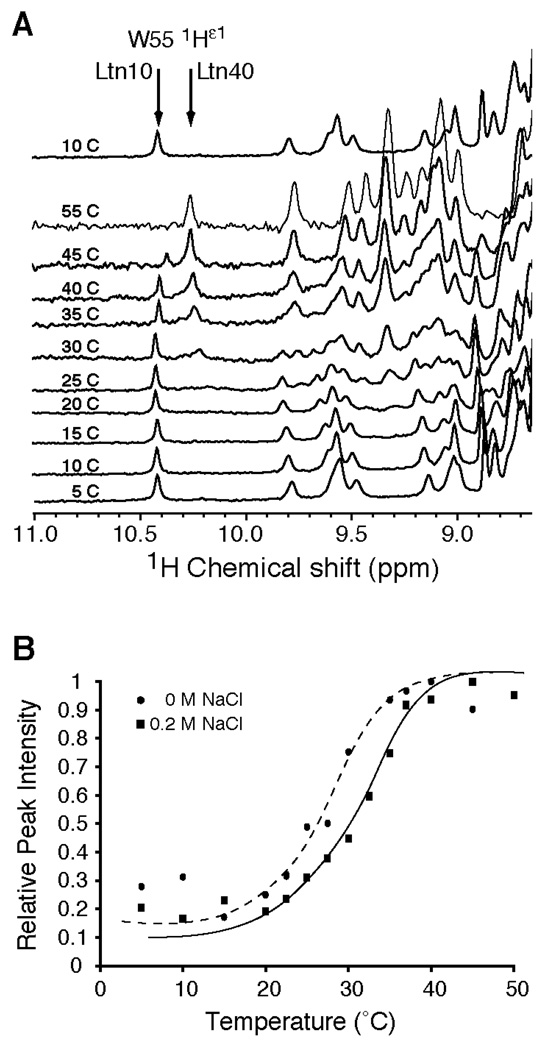
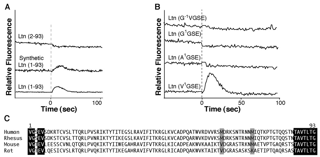
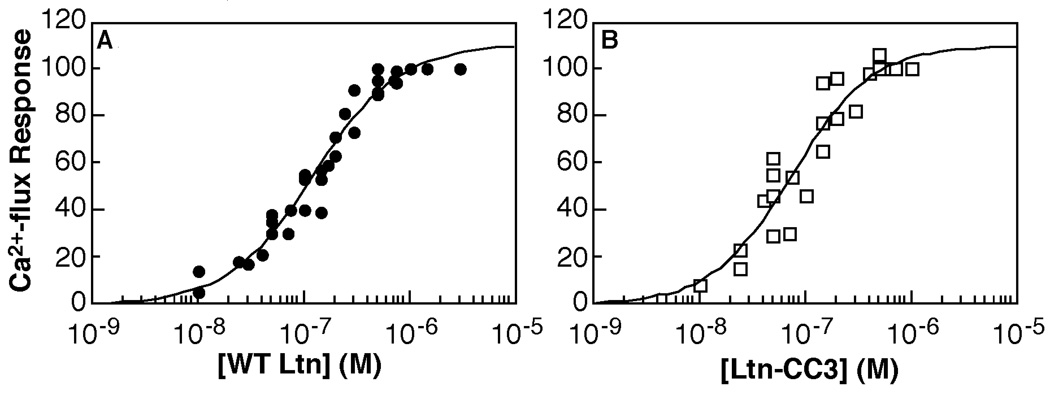
Chemokines adopt a conserved tertiary structure stabilized by two disulfide bridges and direct the migration of leukocytes. Lymphotactin (Ltn) is a unique chemokine in that it contains only one disulfide and exhibits large-scale structural heterogeneity. Under physiological solution conditions (37 degrees C and 150 mM NaCl), Ltn is in equilibrium between the canonical chemokine fold (Ltn10) and a distinct four-stranded beta-sheet (Ltn40). Consequently, it has not been possible to address the biological significance of each structural species independently. To stabilize the Ltn10 structure in a manner independent of specific solution conditions, Ltn variants containing a second disulfide bridge were designed. Placement of the new cysteines was based on a sequence alignment of Ltn with either the first (Ltn-CC1) or third disulfide (Ltn-CC3) in the CC chemokine, HCC-2. NMR data demonstrate that both CC1 and CC3 retain the Ltn10 chemokine structure and no longer exhibit structural rearrangement. The ability of each mutant to activate the Ltn receptor, XCR1, has been tested using an intracellular Ca2+ flux assay. These data support the conclusion that the chemokine fold of Ltn10 is responsible for receptor activation. We also examined the role of amino- and carboxyl-terminal residues in Ltn-mediated receptor activation. In contrast to previous reports, we find that the 25 residues comprising the novel C-terminal extension do not participate in receptor activation, while the native N-terminus is absolutely required for Ltn function.


Figures

Figure 1
Figure 1
Ltn exhibits a reversible structural rearrangement between two distinct structural species. A. 1D-1H NMR spectra of wild-type Ltn (200 µM in 20 mM sodium phosphate, 200mM NaCl, pH 6.0) were acquired at various temperatures. During the Ltn10 to Ltn40 transition, Trp-55 moves from a buried position in the hydrophobic core of Ltn10 to a solvent exposed, unstructured region of Ltn40, which reflected in the chemical shift value for the Nε-1H from 10.2 ppm to 10.4 ppm for the Ltn10 and Ltn40 structures, respectively. The fractional population of Ltn10 and Ltn40 species can be assessed from the intensity of the 1H-Nε peak at 10.2 versus 10.4 ppm. To demonstrate the reversibility of the conformational rearrangement, a spectrum reacquired at 10 °C following the temperature titration is illustrated after the 55 °C spectrum. B. A plot of 1H-Nε peak intensity at 10.4 ppm versus temperature was conducted using wild-type Ltn in the absence (closed circles and dashed trendline) or presence (closed squares and solid trendline) of 200mM NaCl. Peak intensities have been normalized to the maximal intensity obtained after conversion to Ltn40 species. Trendlines were visually fit to the data to represent the shift in relative peak intensity after inclusion of salt to the protein sample.
Figure 2
Figure 2
Introduction of a second disulfide bond in Ltn stabilizes the Ltn10 structure. A. Placement of a second disulfide bond was based on alignment of Ltn with the CC chemokine, HCC-2. Construction of Ltn-CC1 (T10C/iG32ACS33) is based on alignment of the first disulfide in HCC-2, while the position of cysteine substitutions in Ltn-CC3 (V21C/V59C) are based on the unusual third disulfide present in HCC-2. B. 1H-15N HSQC spectra acquired for CC1 and CC3 at 40°C are compared with those taken for wild-type Ltn at 10, 37, and 40°C. Spectra for wild-type protein at 10 and 37°C were acquired in 20mM sodium phosphate, pH 6.0 containing 200 mM NaCl. Spectra of all other protein samples were acquired using 20mM sodium phosphate, pH 6.0 in the absence of salt. HSQC spectra of CC1 and CC3 Ltn resemble the patterns observed for Ltn10. Neither mutant displays evidence for a mixture of the two structural species as observed for the wild-type protein at 37°C. The 1HN peak for Val56 is boxed in the WT spectra acquired at 10°C and the CC1 and CC3 mutants. Owing to ring-current effects of W55, the proton chemical shift for Val56 is significantly upfield of other backbone amide protons, and is indicative of the conserved chemokine fold. C. Amide chemical shifts for the CC1 and CC3 mutants are plotted against the corresponding values for wild-type Ltn at 10 and 40 °C. The excellent correlation of the Ltn10 chemical shifts versus the disulfide-stabilized mutants as compared to the poor correlation with the Ltn40 chemical shifts, indicate that CC1 and CC3 adopt the chemokine-like fold. Outliers in each plot are labeled and correspond to residues at or adjacent to the substituted position in the amino acid sequence.
Figure 3
Figure 3
Solution structure for Ltn-CC3, demonstrates Ltn-CC3 adopts the Ltn10 fold. A. Ensemble of 20 conformers for CC3 (residues 9–75) is shown in Cα trace and rotated 180°about the y-axis. Disulfides are highlighted in yellow. B. Backbone Cα r.m.s.d. values and C. 1H-15N heteronuclear NOEs are plotted for each residue of CC3. D. Overlay of wild-type Ltn10 (cyan) and CC3 (magenta) structures (residues 9–68), rotated 90°about the y-axis. The r.s.m.d. difference for Cα trace is 1.55 Å between Ltn10 and CC3 for residues 15–64.
Figure 4
Figure 4
Ltn requires a native N-terminus for activation of XCR1. A. Comparison of Ca2+ flux response using Ltn(1–93) obtained from a commercial source (R&D Systems) or factor Xa recombinant Ltn versus His8-TEV recombinant Ltn(2–93). Addition of Ltn is indicated with dashed line. All protein concentrations were 100 nM. B. Ca2+ flux response of recombinant Ltn proteins containing either a +1 N-terminal sequence (G−1V1GSE-Ltn) or a substitution of the N-terminal valine with either a glycine or alanine are compared with native N-terminal, Ltn(1–93) obtained from cyanogen bromide cleavage of recombinant protein. All protein concentrations are 500 nM. C. Alignment of Ltn protein sequences from various species indicates that the N-terminus exhibits more sequence variability compared to the C-terminus of Ltn orthologs. To accommodate CnBr treatment, methionine residues at position 63 and 72 were substituted based on sequence alignments with other Ltn proteins. Neither methionine is absolutely conserved, therefore, an M63V and M72A double mutant was generated.
Figure 5
Figure 5
Ltn-CC3 is an XCR1 agonist. CC3 (panel B) activates XCR1 with a similar EC50 value (75.8 ± 17.4 nM) as WT (panel A) (EC50 = 128.1 ± 17.0 nM) by in vitro Ca2+ flux assay. Both proteins consist of residues 1–93 and contain the M63V/M72A substitutions. Complete data from three independent data sets for wild-type and two independent data sets for CC3 were fit to determine EC50 values. Data were normalized to carbachol control as described in experimental procedures and plotted as a percent of maximum response.
Figure 6
Figure 6
The unstructured dynamically disordered C-terminus of Ltn is not required for activation of XCR1. A. Recombinant Ltn proteins containing C-terminal truncations were constructed on the Ltn(M63V/M72A) background. Protein purity and expected molecular weights for each truncation mutant were verified by MALDI-MS. B. Recombinant Ltn proteins containing C-terminal truncations were tested for their ability to induce Ca2+ flux response. Addition of chemokine is indicated with dashed line. All protein concentrations were 200nM. Only Ltn(1–53), which can not adopt the Ltn10 structure did not activate the Ltn receptor. C. Comparison of 1H-15N HSQC spectra for wild-type Ltn and Ltn(1–72) proteins obtained at 10°C in 20mM sodium phosphate buffer, pH 6.0 containing 200 mM NaCl indicate that truncation of last 22 residues does not affect maintenance of Ltn10 structure. D. Comparison of 1H-15N HSQC spectra for wild-type Ltn and Ltn(1–53) proteins obtained at 40°C in 20mM sodium phosphate buffer, pH 6.0. While Ltn(1–53) does not obtain the chemokine fold, it does maintain some characteristics of the Ltn40 structure.

References

    1. Zlotnik A, Yoshie O. Chemokines: a new classification system and their role in immunity. Immunity. 2000;12:121–127. - PubMed
    1. Kelner G, Kennedy J, Bacon K, Kleyensteuber S, Largaespada D, Jenkins N, Copeland N, Bazan J, Moore K, Schall T, Zlotnik A. Lymphotactin: a cytokine that represents a new class of chemokine. Science. 1994;266:1395–1399. - PubMed
    1. Kuloglu ES, McCaslin DR, Markley JL, Volkman BF. Structural rearrangement of human lymphotactin, a C chemokine, under physiological solution conditions. J Biol Chem. 2002;277:17863–17870. - PMC - PubMed
    1. Kuloglu ES, McCaslin DR, Kitabwalla M, Pauza CD, Markley JL, Volkman BF. Monomeric Solution Structure of the Prototypical 'C' Chemokine Lymphotactin. Biochemistry. 2001;40:12486–12496. - PMC - PubMed
    1. Yoshida T, Imai T, Kakizaki M, Nishimura M, Takagi S, Yoshie O. Identification of single C motif-1/lymphotactin receptor XCR1. J Biol Chem. 1998;273:16551–16554. - PubMed

Publication types