. 2007 Jun 1;404(2):197-206.
doi: 10.1042/BJ20061297.

Regulation of oncogenic transcription factor hTAF(II)68-TEC activity by human glyceraldehyde-3-phosphate dehydrogenase (GAPDH)

Affiliations

Regulation of oncogenic transcription factor hTAF(II)68-TEC activity by human glyceraldehyde-3-phosphate dehydrogenase (GAPDH)

Sol Kim et al. Biochem J. .

Abstract

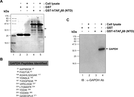
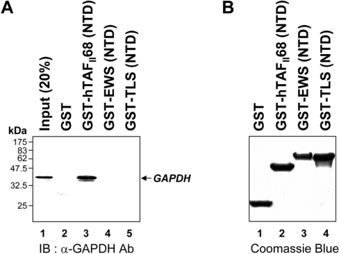
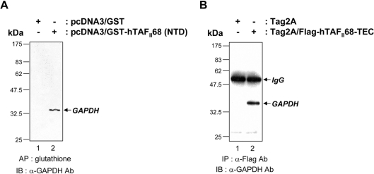
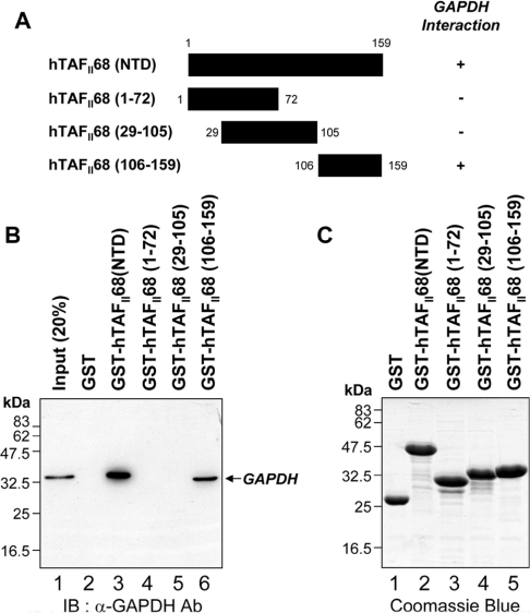
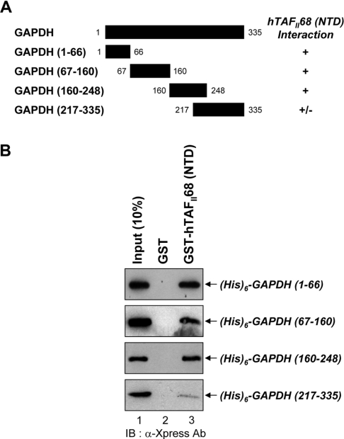
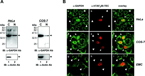
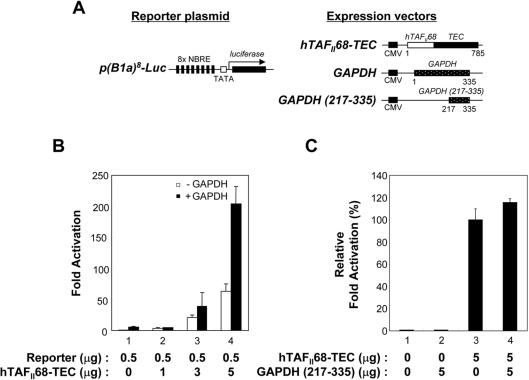
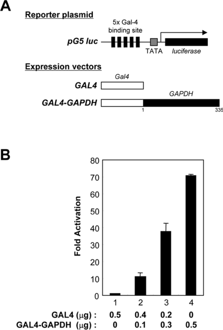
Tumour-specific chromosomal rearrangements are known to create chimaeric products with the ability to generate many human cancers. hTAF(II)68-TEC (where hTAF(II)68 is human TATA-binding protein-associated factor II 68 and TEC is translocated in extraskeletal chondrosarcoma) is such a fusion product, resulting from a t(9;17) chromosomal translocation found in extraskeletal myxoid chondrosarcomas, where the hTAF(II)68 NTD (N-terminal domain) is fused to TEC protein. To identify proteins that control hTAF(II)68-TEC function, we used affinity chromatography on immobilized hTAF(II)68 (NTD) and MALDI-TOF (matrix-assisted laser-desorption ionization-time-of-flight) MS and isolated a novel hTAF(II)68-TEC-interacting protein, GAPDH (glyceraldehyde-3-phosphate dehydrogenase). GAPDH is a glycolytic enzyme that is also involved in the early steps of apoptosis, nuclear tRNA export, DNA replication, DNA repair and transcription. hTAF(II)68-TEC and GAPDH were co-immunoprecipitated from cell extracts, and glutathione S-transferase pull-down assays revealed that the C-terminus of hTAF(II)68 (NTD) was required for interaction with GAPDH. In addition, three independent regions of GAPDH (amino acids 1-66, 67-160 and 160-248) were involved in binding to hTAF(II)68 (NTD). hTAF(II)68-TEC-dependent transcription was enhanced by GAPDH, but not by a GAPDH mutant defective in hTAF(II)68-TEC binding. Moreover, a fusion of GAPDH with the GAL4 DNA-binding domain increased the promoter activity of a reporter containing GAL4 DNA-binding sites, demonstrating the presence of a transactivation domain(s) in GAPDH. The results of the present study suggest that the transactivation potential of the hTAF(II)68-TEC oncogene product is positively modulated by GAPDH.


Figures

Figure 1
Figure 1. Identification of GAPDH as an hTAFII68-TEC-associated protein
(A) SDS/PAGE of hTAFII68 (NTD) complexes isolated from HEK-293T cells. Total HEK-293T cell lysates were incubated with either GST or a GST-fusion protein containing the NTD of hTAFII68 (indicated above the panel). Complexes were resolved by SDS/PAGE (15% gels) and stained with Coomassie Blue. Molecular mass markers are shown on the left; they are derived from prestained protein standards (broad range; New England Biolabs). The band investigated in this analysis is indicated by the arrow to the right. Lane 1, molecular mass marker; lane 2, GST; lane 3, GST plus cell lysate; lane 4, GST–hTAFII68 (NTD); lane 5, GST–hTAFII68 (NTD) plus cell lysate. (B) Peptide sequences of GAPDH identified by MALDI-TOF analysis. The GAPDH peptides matched by MALDI-TOF are shown with amino acid numbers displayed at both ends. (C) Association of hTAFII68 (NTD) with GAPDH. HEK-293T cell lysate was incubated with either GST–hTAFII68 (NTD) or GST. After affinity-selection, the pellets from GST pull-down assays were analysed by SDS/PAGE (15% gels), and bound GAPDH was detected with anti-GAPDH antibody (MAB374; Chemicon) and chemiluminescence (Perkin Elmer Life Science). The positions of the molecular mass markers are indicated on the left-hand side, and the GAPDH band is indicated by the arrow on the right-hand side. Lane 1, GST; lane 2, GST plus cell lysate; lane 3, GST–hTAFII68 (NTD); lane 4, GST–hTAFII68 (NTD) plus cell lysate. IB, immunoblotting; Ab, antibody.
Figure 2
Figure 2. Binding of GAPDH to hTAFII68 (NTD) in vitro
(A) Specific association of hTAFII68 (NTD) with GAPDH. GST-fusion proteins containing the NTDs of hTAFII68, EWS or TLS were incubated with cell lysates. An aliquot of the inputs (20%) and the pellets from the various pull-downs were analysed on SDS/PAGE (15% gels) and bound GAPDH protein was detected using an anti-GAPDH antibody (MAB374). The identities of the GST-fusion proteins are indicated above the panel. The positions of the molecular mass markers are indicated on the left-hand side. GAPDH is indicated by an arrow on the right-hand side. Three independent experiments were performed, all of which gave similar results. Lane 1, 20% input; lane 2, GST alone; lane 3, GST–hTAFII68 (NTD); lane 4, GST–EWS (NTD); lane 5, GST–TLS (NTD). IB, immunoblotting; Ab, antibody. (B) Quantitation of the GST-fusion proteins used in the GST pull-down assays. The GST-fusion proteins utilized in the pull-down assays were fractionated by SDS/PAGE (15% gels) and visualized by Coomassie Blue staining. Three independent experiments were performed, all of which gave similar results. Lane 1, GST alone; lane 2, GST–hTAFII68 (NTD); lane 3, GST–EWS (NTD); lane 4, GST–TLS (NTD).
Figure 3
Figure 3. Association of hTAFII68-TEC with GAPDH in vivo
(A) Co-affinity purification of hTAFII68 (NTD) with GAPDH from COS-7 cells. After transfection (48 h) of COS-7 cells with 10 μg of either pcDNA3/GST or pcDNA3/GST–hTAFII68 (NTD), cell extracts were prepared as described in the Materials and methods section and affinity-precipitated with glutathione–Sepharose beads. After fractionation by SDS/PAGE (15% gels), the proteins were analysed by Western blot with an anti-GAPDH antibody (MAB374). The identities of the transfected DNAs are indicated above the panel. The positions of the molecular mass markers are indicated on the left-hand side and the position of GAPDH is indicated by the arrow on the right-hand side. Three independent experiments were performed, all of which gave similar results. AP, affinity precipitation; IB, immunoblotting; Ab, antibody. (B) Co-immunoprecipitation of hTAFII68-TEC and GAPDH in vivo. After transfection (48 h) of HeLa cells with 10 μg of either Tag2A or Tag2A/Flag-hTAFII68-TEC, cell extracts were immunoprecipitated with an anti-Flag antibody (M2), resolved by SDS/PAGE (15% gels), and probed with an anti-GAPDH antibody (MAB374). The identities of the transfected DNAs are indicated above the panel. The positions of the molecular mass markers are indicated on the left-hand side, and the positions of IgG and GAPDH are indicated by arrows on the right-hand side. Three independent experiments were performed, all of which gave similar results. IP, immunoprecipitation; IB, immunoblotting; Ab, antibody.
Figure 4
Figure 4. Mapping the hTAFII68 (NTD) region that interacts with GAPDH
(A) Schematic representation of the GST–hTAFII68 (NTD) fusion proteins and their ability to bind to GAPDH. Numbers refer to amino acid residues, and binding ability is indicated by + or −. (B) Strong binding of GAPDH to GST–hTAFII68 (106–159). Recombinant GST–hTAFII68 (NTD) deletion mutants were incubated with HEK-293T cell lysates. Following GST pull-down assays, the bound proteins were eluted with SDS loading buffer and analysed by Western blotting with an anti-GAPDH antibody. The positions of molecular mass markers and of GAPDH are indicated. Three independent experiments were performed, all of which gave similar results. Lane 1, 20% input; lane 2, GST alone; lane 3, GST–hTAFII68 (NTD); lane 4, GST–hTAFII68 (1–72); lane 5, GST–hTAFII68 (29–105); lane 6, GST–hTAFII68 (106–159). IB, immunoblotting; Ab, antibody. (C) Coomassie Blue staining of the GST–hTAFII68 deletions. The amounts of GST-fusion proteins utilized in these assays were fractionated on SDS/PAGE (15% gels) and visualized by Coomassie Blue staining. Lane 1, GST alone; lane 2, GST–hTAFII68 (NTD); lane 3, GST–hTAFII68 (1–72); lane 4, GST–hTAFII68 (29–105); lane 5, GST–hTAFII68 (106–159).
Figure 5
Figure 5. Involvement of at least three independent domains of GAPDH in the interaction with hTAFII68 (NTD)
(A) Schematic representation of the deletion mutants of GAPDH and their ability to bind to hTAFII68 (NTD). Numbers refer to amino acid residues, and binding ability is indicated by + or −. (B) Binding of hTAFII68 (NTD) to GAPDH (1–66), GAPDH (67–160), GAPDH (160–248) and GAPDH (217–335). Deletion mutants of GAPDH containing six histidines were expressed in E. coli, purified with Ni2+-NTA–agarose resin, and incubated with either GST or GST-fusion hTAFII68 (NTD) bound to glutathione–Sepharose beads. An aliquot of the input and the pellets from the GST pull-down assays were analysed by SDS/PAGE (15% gels), and bound GAPDH was detected by Western blotting. The positions of the GAPDH deletion mutants are indicated with arrows on the right-hand side. Three independent experiments were performed, all of which gave similar results. Lane 1, 10% input; lane 2, GST alone; lane 3, GST–hTAFII68 (NTD). IB, immunoblotting; Ab, antibody.
Figure 6
Figure 6. Co-localization of hTAFII68-TEC and GAPDH
(A) Subcellular localization of GAPDH in HeLa and COS-7 cells. To determine the subcellular location of GAPDH, HeLa and COS-7 cells were fractionated into nuclear and cytoplasmic preparations. Proteins were separated by SDS/PAGE (15% gels) and detected by Western blotting using anti-GAPDH (MAB 374, upper panels) or anti-actin (I-19, lower panels) antibodies. Three independent experiments were performed, all of which gave similar results. IB, immunoblotting; Ab, antibody. (B) Subcellular distribution of hTAFII68-TEC and GAPDH. HeLa, COS-7, and human chondrocyte cells (C28/I2) grown on coverslips were transfected with a mammalian expression vector encoding Flag-tagged hTAFII68-TEC protein. The transiently transfected cells were fixed with an acetone/methanol mixture and incubated with primary antibodies for GAPDH (V-18) or Flag tag (M2). The subcellular distribution of GAPDH or hTAFII68-TEC was examined using a confocal laser scanning microscope (LSM5 Pascal, Carl Zeiss Co., Ltd.). The merged image (overlay) shows the co-localization. Three independent experiments were performed, all of which gave similar results.
Figure 7
Figure 7. GAPDH enhances hTAFII68-TEC-mediated transcription
(A) Schematic representation of the reporter and expression plasmids used in the present study. The p(B1a)8-Luc reporter plasmid contains eight copies of NBRE upstream of a basal promoter-luciferase gene construct. The eight copies of hTAFII68-TEC recognition sites are indicated by solid bars, the TATA box is represented by an open box, and the luciferase gene is indicated by a solid box. The expression vectors driving the production of hTAFII68-TEC, GAPDH or GAPDH (217–335) are also shown. The positions of the first and last amino acids are indicated below each construct. CMV, cytomegalovirus promoter. (B) Stimulation of hTAFII68-TEC-mediated transactivation by GAPDH. Aliquots of hTAFII68-TEC (bars 1–4) were co-transfected into HeLa cells with 2 μg of empty vector (open bars) or GAPDH expression plasmid (black bars). After 48 h the cells were harvested and luciferase assays performed. The experiments were repeated three times, and the averages of two independent experiments are presented and error bars are shown. (C) A GAPDH (217–335) mutant lacking the hTAFII68-TEC-interacting domain does not stimulate hTAFII68-TEC-mediated transcriptional activation. HeLa cells were transfected with 1 μg of p(B1a)8-Luc reporter plasmid, 5 μg of hTAFII68-TEC, and 5 μg of GAPDH (217–335) or empty vector, and luciferase activity was assayed. Relative transcriptional activation values were calculated with hTAFII68-TEC taken as 100%. The experiments were repeated three times, and the average of two independent experiments are presented with error bars.
Figure 8
Figure 8. Transactivation by a GAL4–GAPDH fusion protein
(A) Schematic representation of the reporter plasmid and expression vectors. The pG5 luc reporter plasmid contains five GAL4-binding sites upstream of the luciferase gene. The GAL4 recognition sites are indicated with solid bars, the TATA box is represented by a shaded box, and the luciferase gene by a solid box. The expression vectors driving the production of GAL4 or GAL4–GAPDH are also presented. The positions of the first and last amino acids of the GAPDH construct are indicated below the construct. (B) Transactivation by GAPDH. COS-7 cells were co-transfected with 0.1 μg of pG5 luc reporter plasmid and 0, 0.1, 0.3 or 0.5 μg of GAL4–GAPDH (bars 1–4). For all reactions the total amount of transfected pGAL4–GAPDH DNA was adjusted to 0.5 μg with empty vector, pGAL4. After 48 h the cells were harvested and luciferase assays were performed. The experiments were repeated three times, and the average of two independent experiments is presented and the error bars are shown.

References

    1. Clark J., Benjamin H., Gill S., Sidhar S., Goodwin G., Crew J., Gusterson B. A., Shipley J., Cooper C. S. Fusion of the EWS gene to CHN, a member of the steroid/thyroid receptor gene superfamily, in a human myxoid chondrosarcoma. Oncogene. 1996;12:229–235. - PubMed
    1. Labelle Y., Zucman J., Stenman G., Kindblom L. G., Knight J., Turc-Carel C., Dockhorn-Dworniczak B., Mandahl N., Desmaze C., Peter M., et al. Oncogenic conversion of a novel orphan nuclear receptor by chromosome translocation. Hum. Mol. Genet. 1995;4:2219–2226. - PubMed
    1. Labelle Y., Bussieres J., Courjal F., Goldring M. B. The EWS/TEC fusion protein encoded by the t(9;22) chromosomal translocation in human chondrosarcomas is a highly potent transcriptional activator. Oncogene. 1999;18:3303–3308. - PubMed
    1. Sjogren H., Meis-Kindblom J., Kindblom L. G., Aman P., Stenman G. Fusion of the EWS-related gene TAF2N to TEC in extraskeletal myxoid chondrosarcoma. Cancer Res. 1999;59:5064–5067. - PubMed
    1. Attwooll C., Tariq M., Harris M., Coyne J. D., Telford N., Varley J. M. Identification of a novel fusion gene involving hTAFII68 and CHN from a t(9;17)(q22;q11.2) translocation in an extraskeletal myxoid chondrosarcoma. Oncogene. 1999;18:7599–7601. - PubMed

Publication types

MeSH terms

Substances