. 2007 May;186(1):150-5.
doi: 10.1016/j.jmr.2007.01.017. Epub 2007 Jan 30.

Towards hyperpolarized (13)C-succinate imaging of brain cancer

Affiliations

Towards hyperpolarized (13)C-succinate imaging of brain cancer

Pratip Bhattacharya et al. J Magn Reson. 2007 May.

Abstract

We describe a novel (13)C enriched precursor molecule, sodium 1-(13)C acetylenedicarboxylate, which after hydrogenation by PASADENA (Parahydrogen and Synthesis Allows Dramatically Enhanced Nuclear Alignment) under controlled experimental conditions, becomes hyperpolarized (13)C sodium succinate. Fast in vivo 3D FIESTA MR imaging demonstrated that, following carotid arterial injection, the hyperpolarized (13)C-succinate appeared in the head and cerebral circulation of normal and tumor-bearing rats. At this time, no in vivo hyperpolarized signal has been localized to normal brain or brain tumor. On the other hand, ex vivo samples of brain harvested from rats bearing a 9L brain tumor, 1 h or more following in vivo carotid injection of hyperpolarized (13)C sodium succinate, contained significant concentrations of the injected substrate, (13)C sodium succinate, together with (13)C maleate and succinate metabolites 1-(13)C-glutamate, 5-(13)C-glutamate, 1-(13)C-glutamine and 5-(13)C-glutamine. The (13)C substrates and products were below the limits of NMR detection in ex vivo samples of normal brain consistent with an intact blood-brain barrier. These ex vivo results indicate that hyperpolarized (13)C sodium succinate may become a useful tool for rapid in vivo identification of brain tumors, providing novel biomarkers in (13)C MR spectral-spatial images.


Figures
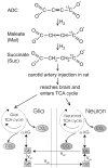
Fig. 1
Fig. 1
A representation of intra-cerebral metabolite cycling between neurons and glia believed to underlie glutamate neurotransmission. The diagram of experimental design is shown above. Twenty-five millimolar aqueous solution of ADC precursor is hydrogenated in the polarizer to yield 3 mL of maleate and succinate products, which is then injected in the carotid artery of 9L tumor bearing rat. We propose that the hydrogenated products reach the brain through an altered blood–brain barrier in tumor tissue and enter glial and neuronal TCA cycle to yield glutamine and glutamate as final product in vivo.
Fig. 2
Fig. 2
(a) 13C NMR spectrum of hyperpolarized 1-13C-succinate from ADC precursor acquired at 4.7 T Bruker horizontal wide-bore MR system. A sphere containing 2.8 mL of 4.4 M 1-13C-acetate solution is used as a reference at ~182 ppm. A single transient 13C NMR spectrum revealed ~4400-fold signal enhancement with respect to Boltzmann polarization. (b) 13C spectrum of the post-hyperpolarized reaction mixture at Boltzmann polarization, 128 transients.
Fig. 3
Fig. 3
In vivo13C 3D FIESTA imaging of rat brain with PASADENA-hyperpolarized succinate. Sub-second 13C images (0.3 s) were acquired using a multi-nuclear 3D fast imaging sequence employing balanced steady-state acquisition (3D FIESTA). To allow for the short lived polarization, the 3D FIESTA 13C data were acquired with a TR/TE of 6.3/3.1 ms, sixteen 5 mm slices, 44 phase encodings over 220 mm field-of-view (FOV) and 64 readout points over 320 mm FOV to yield an isotropic spatial resolution of 5 × 5 × 5 mm3. The image shown represents one slice of 13C data acquired 9 s after infusion. It is overlaid on a coronal 3D fast gradient echo proton image with matching FOV and slice location acquired prior to infusion to provide anatomical correlation. All data were acquired on a 1.5 T (GE LX 9.1, Waukesha, WI) MR scanner with a dual-tuned 1H/13C custom saddle coil. The rat was anesthetized and cannulated in the carotid artery where upon 1.5 mL of 25 mM of hyperpolarized succinate was injected. For reference, a 4.4 M acetate phantom was placed next to the rat, demonstrating significant signal enhancement of hyperpolarized succinate in vivo given the difference in concentration (4.4 M vs. 0.025 M) and signal intensity.
Fig. 4
Fig. 4
Ex vivo MAS 13C spectra of brain (lower) and brain tumor tissues (upper). 80 mg of tissue was used in each experiment. 13C MAS spectra were acquired under slow spinning, 1–2 kHz conditions, low power 1H decoupling and 4 °C utilizing spin echo pulse sequence with t90° = 4.0 μs, 1024 transients and recycling delay of 5 s. Representative in vivo RARE image of 9L tumor bearing brain is shown. 13C precursors, succinate and maleate, as well as putative products of tumor metabolism, glutamine (Gln) and glutamate (Glu) are assigned based on model solutions. Note the absence of 13C enrichment in normal brain tissue from the same animal.

References

    1. Edelman R, Hesselink J, Zlatkin M. Clinical Magnetic Resonance Imaging. W.B. Saunders and Co.; Philadelphia: 1996.
    1. Magnetic Resonance at Solid State Physics and Magnetic Resonance Service in Saclay. Bulletin D Informations Scientifiques Et Techniques. 1969:3.
    1. Modern Techniques in Neuroscience Research. Springer–Verlag; 1999.
    1. Cerdan S. NMR in Biomedicine. Wiley, NY; 2003.
    1. Ross B, Lin A, Harris K, Bhattacharya P, Schweinsburg B. Clinical experience with C-13 MRS in vivo. NMR Biomed. 2003;16:358–369. - PubMed

Publication types