Serial MRI to determine the effect of dexamethasone on the cerebral pathology of tuberculous meningitis: an observational study
- PMID: 17303529
- PMCID: PMC4333204
- DOI: 10.1016/S1474-4422(07)70034-0
Serial MRI to determine the effect of dexamethasone on the cerebral pathology of tuberculous meningitis: an observational study
Abstract
Background: Adjunctive dexamethasone increases survival from tuberculous meningitis, but the underlying mechanism is unclear. We aimed to determine the effect of dexamethasone on cerebral MRI changes and their association with intracerebral inflammatory responses and clinical outcome in adults treated for tuberculous meningitis.
Methods: Cerebral MRI was undertaken, when possible, at diagnosis and after 60 days and 270 days of treatment in adults with tuberculous meningitis admitted to two hospitals in Vietnam. Patients were randomly assigned either dexamethasone (n=24) or placebo (n=19) and received 9 months of treatment with standard first-line antituberculosis drugs. We assessed associations between MRI findings, treatment allocation, and resolution of fever, coma, cerebrospinal fluid inflammation, and neurological outcome.
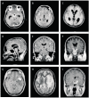
Findings: 83 scans were done for 43 patients: 19 given placebo, 24 given dexamethasone. Basal meningeal enhancement (82%) and hydrocephalus (77%) were the most common presenting findings. Fewer patients had hydrocephalus after 60 days of treatment with dexamethasone than after placebo treatment (p=0.217). Tuberculomas developed in 74% of patients during treatment and in equal proportions in the treatment groups; they were associated with long-term fever, but not relapse or poor clinical outcome. The basal ganglia were the most common site of infarction; the proportion with infarction after 60 days was halved in the dexamethasone group (27%vs 58%, p=0.130).
Interpretation: Dexamethasone may affect outcome from tuberculous meningitis by reducing hydrocephalus and preventing infarction. The effect may have been under-estimated because the most severe patients could not be scanned.
Figures


Comment in
-
Brain teasing effect of dexamethasone.Lancet Neurol. 2007 Mar;6(3):203-4. doi: 10.1016/S1474-4422(07)70041-8. Lancet Neurol. 2007. PMID: 17303522 No abstract available.
References
-
- Thwaites GE, Tran TH. Tuberculous meningitis: many questions, too few answers. Lancet Neurol. 2005;4:160–70. - PubMed
-
- Dastur DK, Manghani DK, Udani PM. Pathology and pathogenetic mechanisms in neurotuberculosis. Radiol Clin North Am. 1995;33:733–52. - PubMed
-
- Thwaites GE, Nguyen DB, Nguyen HD, et al. Dexamethasone for the treatment of tuberculous meningitis in adolescents and adults. N Engl J Med. 2004;351:1741–51. - PubMed
-
- Simmons CP, Thwaites GE, Quyen NT, et al. The clinical benefit of adjunctive dexamethasone in tuberculous meningitis is not associated with measurable attenuation of peripheral or local immune responses. J Immunol. 2005;175:579–90. - PubMed
-
- Schoeman JF, Van Zyl LE, Laubscher JA, Donald PR. Effect of corticosteroids on intracranial pressure, computed tomographic findings, and clinical outcome in young children with tuberculous meningitis. Pediatrics. 1997;99:226–31. - PubMed
Publication types
MeSH terms
Substances
Grants and funding
LinkOut - more resources
Full Text Sources
Other Literature Sources
Medical

