. 2007 Mar 7;26(5):1268-78.
doi: 10.1038/sj.emboj.7601583. Epub 2007 Feb 15.

The role of fibroblast growth factor receptor 2b in skin homeostasis and cancer development

Affiliations

The role of fibroblast growth factor receptor 2b in skin homeostasis and cancer development

Richard Grose et al. EMBO J. .

Abstract

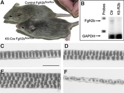
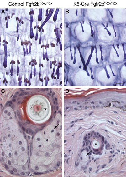
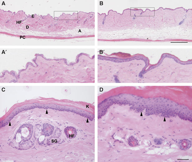
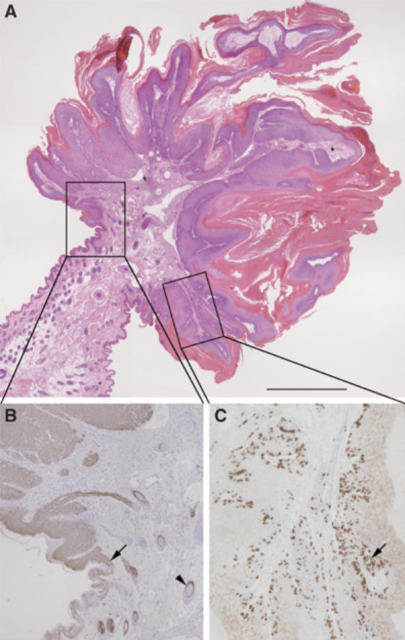
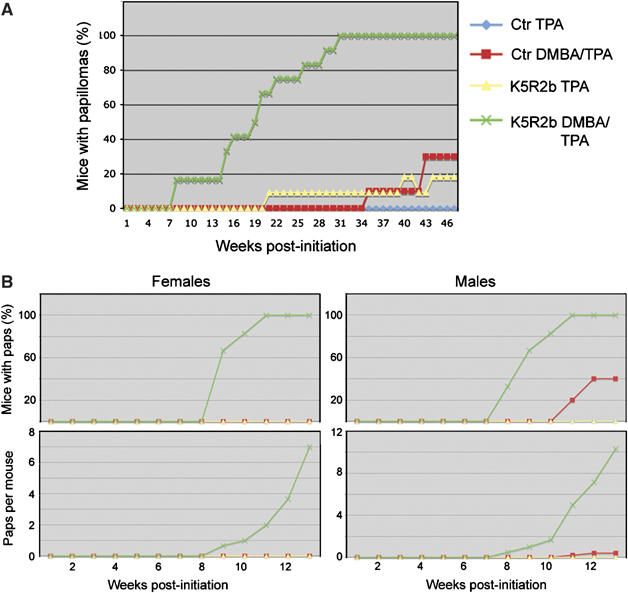
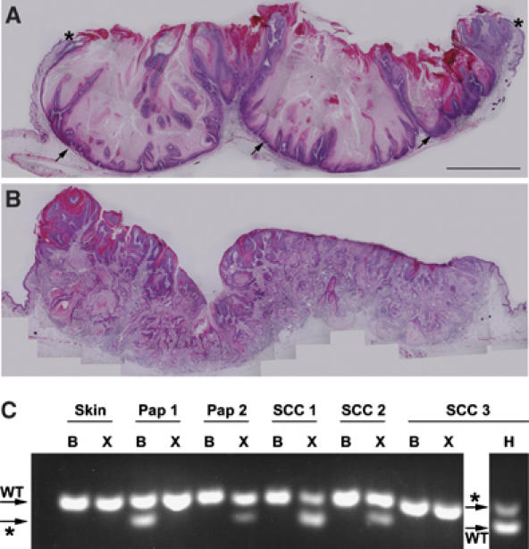
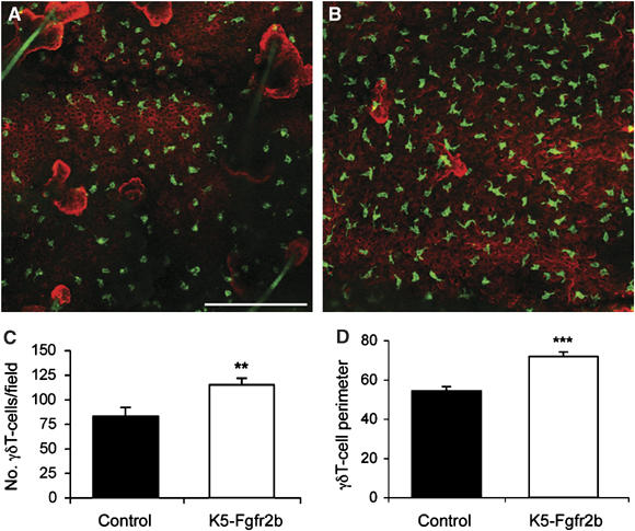
The epithelial isoform of fibroblast growth factor receptor 2 (Fgfr2b) is essential for embryogenesis, and Fgfr2b-null mice die at birth. Using Cre-Lox transgenics to delete Fgfr2b in cells expressing keratin 5, we show that mice lacking epidermal Fgfr2b survive into adulthood but display striking abnormalities in hair and sebaceous gland development. Epidermal hyperthickening develops with age, and 10% of mutant mice develop spontaneous papillomas, demonstrating the role of Fgfr2b in post-natal skin development and in adult skin homeostasis. Mice lacking epithelial Fgfr2b show great sensitivity to chemical carcinogenic insult, displaying several oncogenic ha-ras mutations with dramatic development of papillomas and squamous cell carcinomas. Mutant mice have increased inflammation in the skin, with increased numbers of macrophages and gammadeltaT cells with abnormal morphology. Mutant skin shows several changes in gene expression, including enhanced expression of the pro-inflammatory cytokine interleukin 18 and decreased expression of Serpin a3b, a potential tumor suppressor. Thus we describe a novel role of Fgfr2b and provide the first evidence of a tyrosine kinase receptor playing a tumor suppressive role in the skin.


Figures

Figure 1
Figure 1
Skin-specific deletion of Fgfr2b leads to defective hair formation. (A) Five-week-old male littermates. Both mice are homozygous for the floxed Fgfr2b allele—the lower mouse also carries the K5 Cre transgene. Mice lacking Fgfr2b in the epidermis are normal size compared with wild-type siblings, but show clear coat differences, with their fur appearing sleek and silky compared with wild types. (B) RNase protection assay. Expression of Fgfr2b mRNA in skin from 8-week-old control and K5-R2b-null littermates. Levels of GAPDH mRNA were assayed in the same samples to serve as a control of both loading levels and RNA quality. Each hybridization probe (1000 c.p.m.) was loaded in the lanes labelled ‘probes' and used as size markers. Arrows indicate the protected fragments for Fgfr2b and control (GAPDH) mRNA. (C–F) Pelage hair structure. Light microscopy of the three main hair subtypes from control mice: (C) Zigzag, (D) Guard and (E) Awl hairs. (F) Disorganized zigzag hair from a K5-R2b-null mouse. Scale bar, 20 μm.
Figure 2
Figure 2
Sebaceous gland atrophy in K5-R2b-null mice. By 3 months of age, epidermal whole-mount peroxidase staining (sebaceous glands appear brown) combined with Mayer's hemalum counterstain revealed that sebaceous glands in the tail of K5-R2b-null mice were virtually absent (compare (A) with (B)). This absence was confirmed by scanning through H&E-stained serial transverse sections of tail skin, looking for sebocytes (arrows in (C) and (D); asterisks indicate transversely sectioned hair shafts). (C) and (D) represent sections showing the largest proportion of sebaceous tissue encountered in serial transverse sections from tails of wild-type and K5-R2b-null mice, respectively. Scale bar, 200 μm.
Figure 3
Figure 3
Epidermal hyperthickening in the absence of Fgfr2b. Transverse sections through back and tail skin of 18-month-old mice were stained with H&E. Back skin of control (A and A′) and K5-R2b-null mice (B and B′). In wild-type tail skin (C), the basal layer of keratinocytes is clearly visible as a distinct line of nuclei at the dermo-epidermal junction (arrowheads) and anuclear, keratinized squames can be seen sloughing from the surface. By comparison, Fgfr2b-null tail skin (D) shows regions of dysplasia (arrowheads) and parakeratosis. A, adipose layer; D, dermis; E, epidermis; HF, hair follicle; K, keratinized squames; PC, panniculus carnosus; SG, sebaceous gland. Scale bars, 200 μm (A, B) and 50 μm (C, D).
Figure 4
Figure 4
Spontaneous papilloma formation in the absence of Fgfr2b. (A) H&E staining reveals classical exophytic morphology of spontaneous papillomas, with a stalk of relatively normal skin leading to a hyperthickened, keratinized papilloma packed with inflammatory cells. Higher magnification views of the boxed areas in (A) show that papilloma keratinocytes express keratin 6 (brown immunostaining in (B)), normally restricted to hair follicles (arrowhead), from the margin, where the papilloma begins (arrow). BrdU staining for proliferating cells (C) in both basal and suprabasal layers (arrow). Scale bar, 200 μm.
Figure 5
Figure 5
Enhanced sensitivity to skin carcinogenesis in K5-R2b-null mice. Cohorts of female mice were subjected to classical two-step skin carcinogenesis treatment. (A) Female mice were treated with DMBA or solvent acetone alone at 8 weeks of age and then all treated twice weekly with TPA for 15 weeks. In a repeat study (B), the protocol was adjusted slightly, such that male and female mice were treated, giving a total of eight groups (key same as for (A)). The treatment regime differed only in that the mice received once weekly applications of a double dose of TPA.
Figure 6
Figure 6
Progression of skin carcinogenesis lesions to squamous cell carcinoma. Transverse sections through lesions from K5-R2b-null mice highlight keratoacanthoma (A) and squamous cell carcinoma (B). Although keratoacanthomas no longer exhibit the exophytic growth of papillomas, with their margins covered with normal skin (asterisks in (A)), they are still non-invasive, with a clearly defined basal layer of keratinocytes (arrows in (A)). In contrast, the lesions that progress to squamous cell carcinoma (B) show no clearly defined basal surface, but rather a mass of invasive keratinocyte projections and a strong recruitment of inflammatory cells. (C) RFLP analysis of mutations in codon 61 of ha-ras. PCR products derived from skin, papilloma (Pap) or squamous cell carcinoma (SCC) digested with BspHI (B), XbaI (X) and Hpy118II (H) were separated on 4% agarose gels. Bands diagnostic of the wild-type (WT) and mutant (*) alleles are indicated (arrows).
Figure 7
Figure 7
γδT-cell abnormalities in K5-R2b-null epidermis. Whole-mount preparations of ear epidermis from control (A) and K5-R2b-null (B) mice stained with antibodies to γδT cells (green) and keratin 14 (red) highlight the increased γδT-cell density in mutant epidermis (quantitated in (C)). In addition to being present in increased numbers, γδT cells in mutant epidermis display an altered morphology, adopting a more dendritic appearance, reflected by a significantly increased cell perimeter (quantitated in (D)). Scale bar in (A), 50 μm. **P<0.01; ***P<0.001 using the Mann--Whitney test.

References

    1. auf dem Keller U, Huber M, Beyer TA, Kumin A, Siemes C, Braun S, Bugnon P, Mitropoulos V, Johnson DA, Johnson JA, Hohl D, Werner S (2006) Nrf transcription factors in keratinocytes are essential for skin tumor prevention but not for wound healing. Mol Cell Biol 26: 3773–3784 - PMC - PubMed
    1. Balkwill F (2004) Cancer and the chemokine network. Nat Rev Cancer 4: 540–550 - PubMed
    1. Balkwill F, Coussens LM (2004) Cancer: an inflammatory link. Nature 431: 405–406 - PubMed
    1. Bernard-Pierrot I, Ricol D, Cassidy A, Graham A, Elvin P, Caillault A, Lair S, Broet P, Thiery JP, Radvanyi F (2004) Inhibition of human bladder tumour cell growth by fibroblast growth factor receptor 2b is independent of its kinase activity. Involvement of the carboxy-terminal region of the receptor. Oncogene 23: 9201–9211 - PubMed
    1. Brakebusch C, Grose R, Quondamatteo F, Ramirez A, Jorcano JL, Pirro A, Svensson M, Herken R, Sasaki T, Timpl R, Werner S, Fassler R (2000) Skin and hair follicle integrity is crucially dependent on beta 1 integrin expression on keratinocytes. EMBO J 19: 3990–4003 - PMC - PubMed

Publication types

MeSH terms

Substances